
Search Count: 10
 |
Organism: Yersinia entomophaga
Method: ELECTRON MICROSCOPY Release Date: 2019-05-08 Classification: TOXIN |
 |
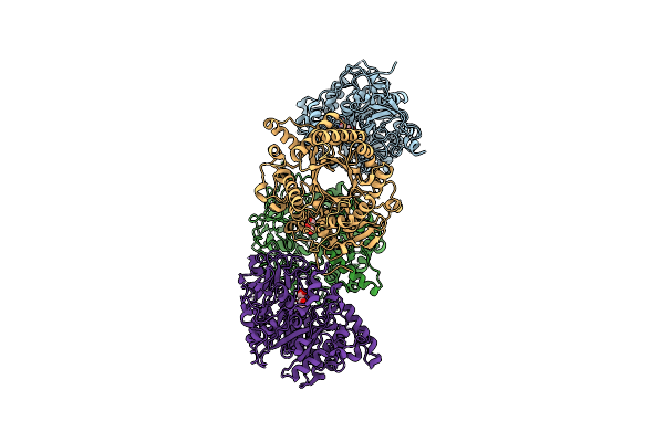
Crystal Structure Of The C-Terminal Toxin Domain Of Rhs2 From Yersinia Entomophaga
Organism: Yersinia entomophaga
Method: X-RAY DIFFRACTION Resolution:1.80 Å Release Date: 2018-08-22 Classification: TOXIN Ligands: PT, CL, BR |
 |
Organism: Yersinia entomophaga
Method: X-RAY DIFFRACTION Resolution:2.40 Å Release Date: 2017-08-09 Classification: TOXIN Ligands: CA, NA, CL |
 |
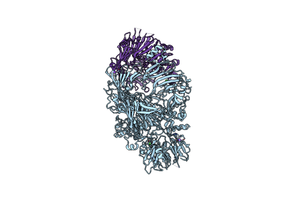
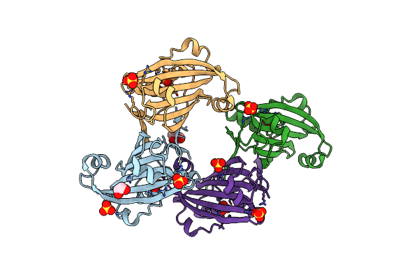
N-Terminal Domain Of Orf22, A Cydia Pomonella Granulovirus Envelope Protein
Organism: Cydia pomonella granulosis virus
Method: X-RAY DIFFRACTION Resolution:1.40 Å Release Date: 2016-03-09 Classification: VIRAL PROTEIN Ligands: ACT |
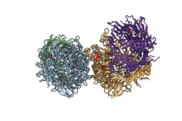 |
Structure Of The Rhs-Repeat Containing Bc Component Of The Secreted Abc Toxin Complex From Yersinia Entomophaga
Organism: Yersinia
Method: X-RAY DIFFRACTION Resolution:2.49 Å Release Date: 2013-08-07 Classification: TOXIN Ligands: GOL, K |
 |
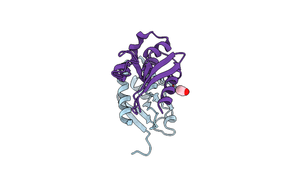
Crystal Structure Of A Chitinase From The Yersinia Entomophaga Toxin Complex
Organism: Yersinia entomophaga
Method: X-RAY DIFFRACTION Resolution:1.80 Å Release Date: 2013-02-27 Classification: SUGAR BINDING PROTEIN Ligands: GOL |
 |
Crystal Structure Of The Chitinase Chi1 Fitted Into The 3D Structure Of The Yersinia Entomophaga Toxin Complex
Organism: Yersinia entomophaga
Method: ELECTRON MICROSCOPY Resolution:17.00 Å Release Date: 2011-11-16 Classification: HYDROLASE |
 |
Organism: Yersinia
Method: X-RAY DIFFRACTION Resolution:1.74 Å Release Date: 2011-08-10 Classification: HYDROLASE Ligands: 2PE, GOL |
 |
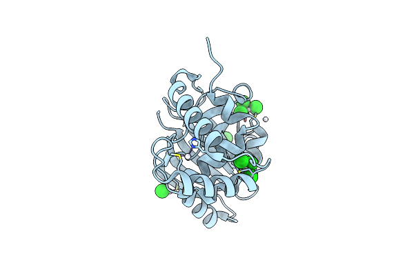
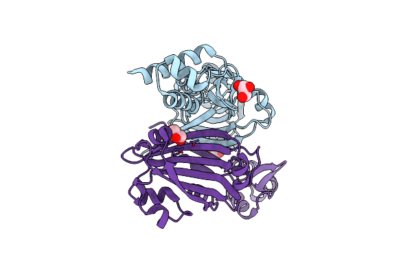
Crystal Structure Of The Effector Domain Of Phnf From Mycobacterium Smegmatis
Organism: Mycobacterium smegmatis
Method: X-RAY DIFFRACTION Resolution:1.90 Å Release Date: 2009-11-17 Classification: TRANSCRIPTION Ligands: SO4, GOL |
 |
Organism: Mycobacterium smegmatis
Method: X-RAY DIFFRACTION Resolution:1.80 Å Release Date: 2009-11-17 Classification: TRANSCRIPTION Ligands: GOL |