
Search Count: 28
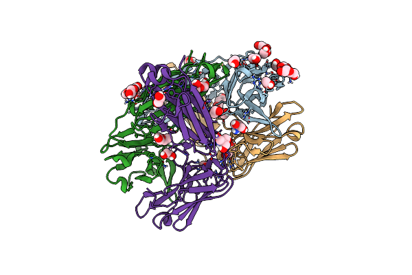 |
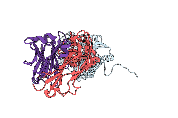
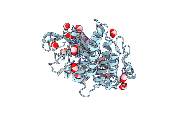
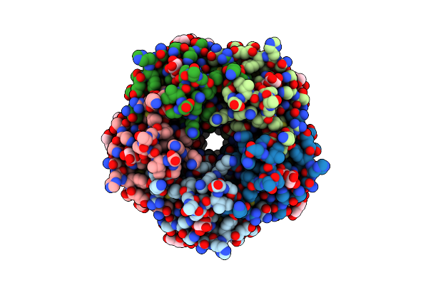
Structure Of The Arginase-2-Inhibitory Human Antigen-Binding Fragment Fab C0021144
Organism: Homo sapiens
Method: X-RAY DIFFRACTION Resolution:2.40 Å Release Date: 2020-06-10 Classification: PROTEIN BINDING Ligands: CL, EDO, PEG, TRS |
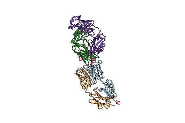 |
Structure Of The Arginase-2-Inhibitory Human Antigen-Binding Fragment Fab C0021158
Organism: Homo sapiens
Method: X-RAY DIFFRACTION Resolution:1.90 Å Release Date: 2020-06-10 Classification: PROTEIN BINDING Ligands: ACT, CL |
 |
Structure Of The Arginase-2-Inhibitory Human Antigen-Binding Fragment Fab C0021181
Organism: Homo sapiens
Method: X-RAY DIFFRACTION Resolution:1.70 Å Release Date: 2020-06-10 Classification: PROTEIN BINDING Ligands: CL, SIN, PEG, PGE |
 |
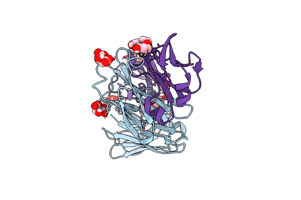
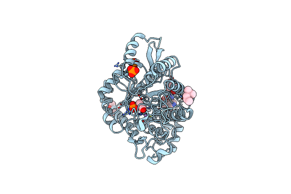
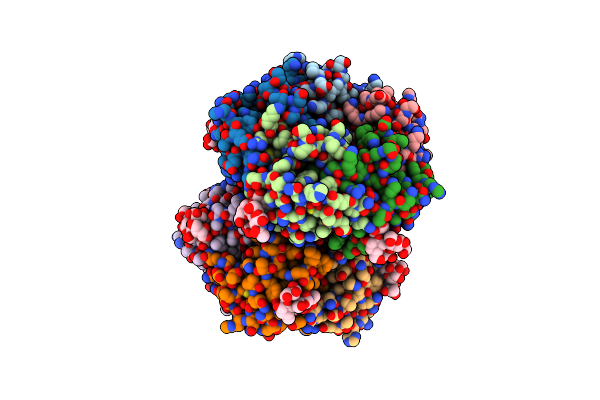
Structure Of Arginase-2 In Complex With The Inhibitory Human Antigen-Binding Fragment Fab C0021158
Organism: Homo sapiens
Method: X-RAY DIFFRACTION Resolution:2.40 Å Release Date: 2020-06-10 Classification: PROTEIN BINDING Ligands: GOL, MN, SO4 |
 |
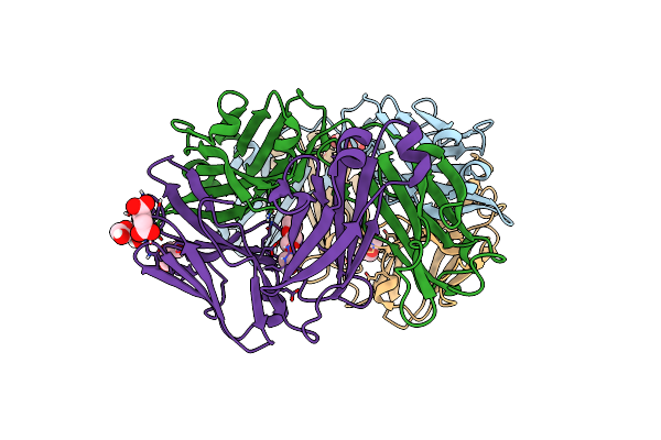
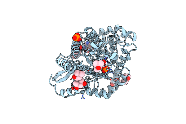
Structure Of Arginase-2 In Complex With The Inhibitory Human Antigen-Binding Fragment Fab C0021181
Organism: Homo sapiens
Method: X-RAY DIFFRACTION Resolution:2.90 Å Release Date: 2020-06-10 Classification: PROTEIN BINDING Ligands: MN, PO4 |
 |
Structure Of The Arginase-2-Inhibitory Human Antigen-Binding Fragment Fab C0020187
Organism: Homo sapiens
Method: X-RAY DIFFRACTION Resolution:1.78 Å Release Date: 2020-06-10 Classification: PROTEIN BINDING Ligands: CL, MPD, EPE, SO4 |
 |
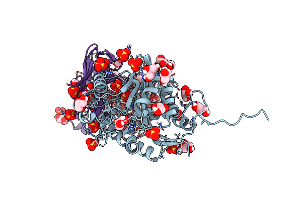
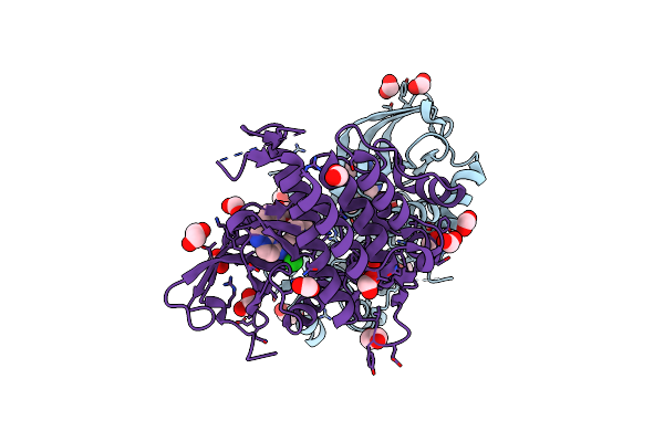
Structure Of Arginase-2 In Complex With The Inhibitory Human Antigen-Binding Fragment Fab C0020187
Organism: Homo sapiens
Method: X-RAY DIFFRACTION Resolution:3.25 Å Release Date: 2020-06-10 Classification: PROTEIN BINDING Ligands: MN, SO4 |
 |
Structure Of The Arginase-2-Inhibitory Human Antigen-Binding Fragment Fab C0021177
Organism: Homo sapiens
Method: X-RAY DIFFRACTION Resolution:2.25 Å Release Date: 2020-06-10 Classification: PROTEIN BINDING Ligands: PG4, P6G, MLT, PGE |
 |
Crystal Structure Of Partially Phosphorylated Ret V804M Tyrosine Kinase Domain Complexed With Pdd00018412
Organism: Homo sapiens
Method: X-RAY DIFFRACTION Resolution:2.05 Å Release Date: 2020-03-11 Classification: ONCOPROTEIN Ligands: H6W, EDO, FMT, CL |
 |
Crystal Structure Of Phosphorylated Ret V804M Tyrosine Kinase Domain Complexed With Pdd00018366
Organism: Homo sapiens
Method: X-RAY DIFFRACTION Resolution:1.88 Å Release Date: 2020-03-11 Classification: ONCOPROTEIN Ligands: H72, FMT |
 |
Organism: Homo sapiens
Method: X-RAY DIFFRACTION Release Date: 2018-08-15 Classification: ANTITUMOR PROTEIN |
 |
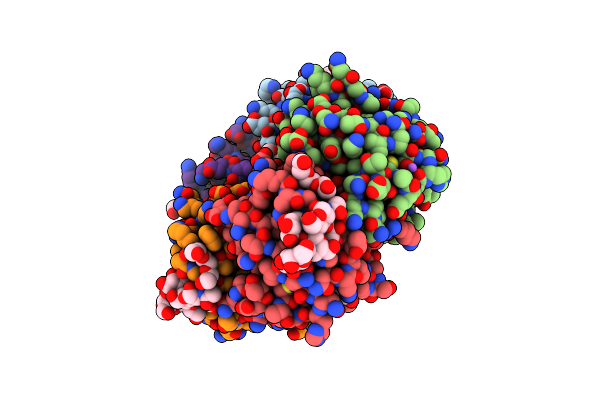
High-Resolution Structure Of Chorismate Mutase From Corynebacterium Glutamicum
Organism: Corynebacterium glutamicum
Method: X-RAY DIFFRACTION Resolution:1.06 Å Release Date: 2017-08-02 Classification: ISOMERASE Ligands: FMT |
 |
Organism: Corynebacterium glutamicum
Method: X-RAY DIFFRACTION Resolution:2.45 Å Release Date: 2017-08-02 Classification: TRANSFERASE Ligands: MN, PEP, MPD, TRP, PO4 |
 |
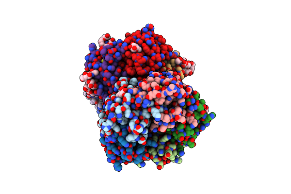
Non-Covalent Complex Of And Dahp Synthase And Chorismate Mutase From Corynebacterium Glutamicum With Bound Transition State Analog
Organism: Corynebacterium glutamicum
Method: X-RAY DIFFRACTION Resolution:2.15 Å Release Date: 2017-08-02 Classification: TRANSFERASE/ISOMERASE Ligands: GOL, PEG, PGE, MN, PO4, TRP, PG4, TSA |
 |
Organism: Corynebacterium glutamicum
Method: X-RAY DIFFRACTION Resolution:2.65 Å Release Date: 2017-08-02 Classification: SUGAR BINDING PROTEIN Ligands: MN, PEP, TRP, MPD, PGE, PO4, TRS |
 |
Organism: Vibrio cholerae o1
Method: X-RAY DIFFRACTION Resolution:1.08 Å Release Date: 2016-03-30 Classification: TOXIN Ligands: CA, GAL, GLA, BCN, PEG |
 |
Organism: Vibrio cholerae o1
Method: X-RAY DIFFRACTION Resolution:1.50 Å Release Date: 2016-03-30 Classification: TOXIN Ligands: CA, BCN, FUC |
 |
Organism: Vibrio cholerae o1
Method: X-RAY DIFFRACTION Resolution:1.40 Å Release Date: 2016-03-30 Classification: TOXIN Ligands: PEG, GAL, NA, MES, PGE, GLA |
 |
Organism: Vibrio cholerae o1
Method: X-RAY DIFFRACTION Resolution:1.60 Å Release Date: 2016-03-30 Classification: TOXIN Ligands: CA, FUC, BCN, 1PE, NAG |
 |
Organism: Vibrio cholerae serotype o1 (strain atcc 39315 / el tor inaba n16961)
Method: X-RAY DIFFRACTION Resolution:1.55 Å Release Date: 2016-03-30 Classification: TOXIN Ligands: CA, BCN, FUC, GLC, PG4 |