
Search Count: 17
 |
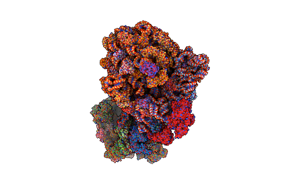
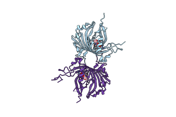
Panoptes Opts Minimal Crispr Polymerase (Mcpol) From Klebsiella Pneumoniae Strain Kp67
Organism: Klebsiella pneumoniae
Method: X-RAY DIFFRACTION Release Date: 2025-09-24 Classification: ANTIVIRAL PROTEIN |
 |
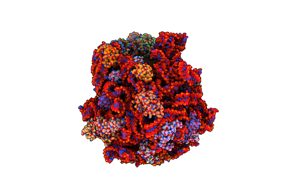
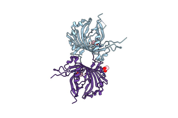
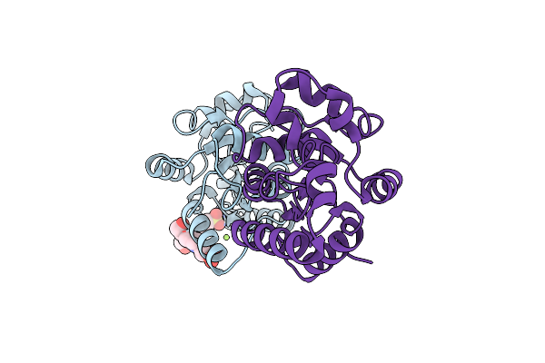
Panoptes Opts Minimal Crispr Polymerase (Mcpol) With Non-Hydrolyzable Ligand Apcpp From Klebsiella Pneumoniae Strain Kp67
Organism: Klebsiella pneumoniae
Method: X-RAY DIFFRACTION Release Date: 2025-09-24 Classification: ANTIVIRAL PROTEIN Ligands: APC, MG |
 |
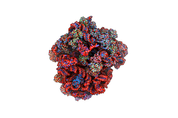
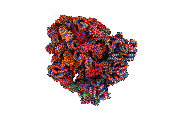
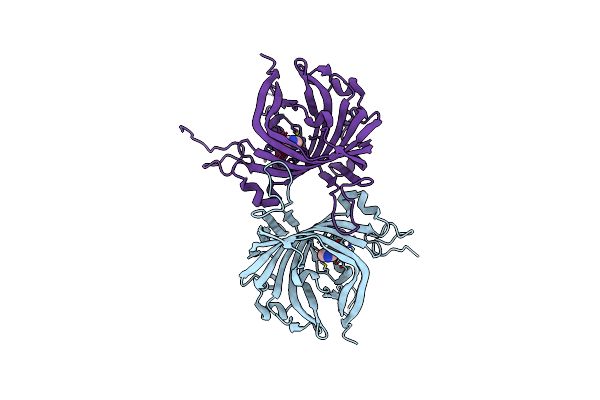
Organism: Escherichia coli, Vibrio alginolyticus
Method: ELECTRON MICROSCOPY Release Date: 2025-02-12 Classification: RIBOSOME |
 |
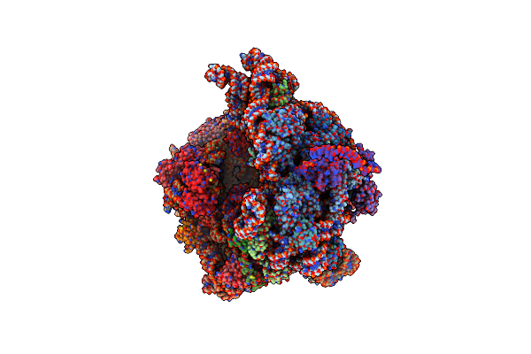
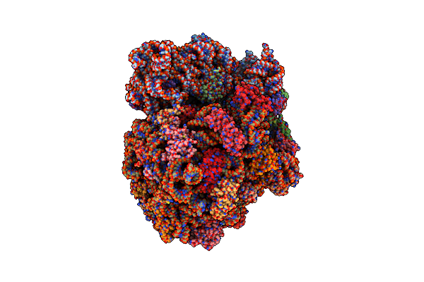
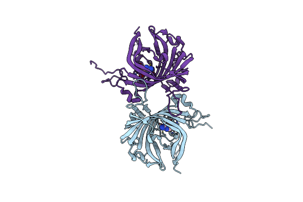
Organism: Escherichia coli, Vibrio alginolyticus
Method: ELECTRON MICROSCOPY Release Date: 2025-02-12 Classification: RIBOSOME |
 |
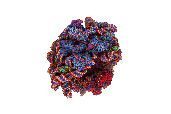
Organism: Bacillus subtilis
Method: ELECTRON MICROSCOPY Release Date: 2024-01-17 Classification: RIBOSOME |
 |
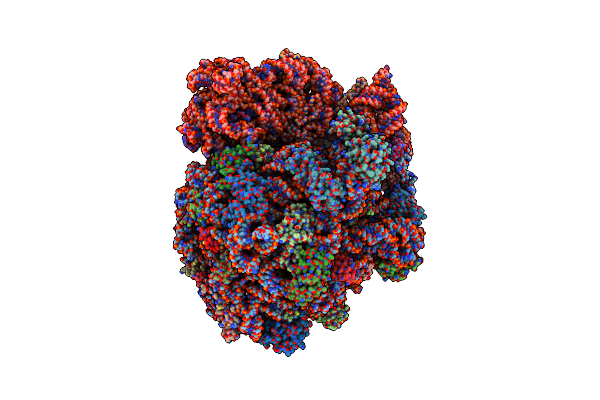
Organism: Bacillus subtilis
Method: ELECTRON MICROSCOPY Release Date: 2023-12-27 Classification: RIBOSOME |
 |
Organism: Escherichia coli, Vibrio alginolyticus
Method: ELECTRON MICROSCOPY Release Date: 2022-06-22 Classification: RIBOSOME Ligands: MG, K |
 |
Organism: Escherichia coli, Vibrio alginolyticus, Escherichia coli k-12, Escherichia coli umea 3162-1
Method: ELECTRON MICROSCOPY Release Date: 2022-04-27 Classification: RIBOSOME Ligands: MG, K |
 |
Organism: Vibrio alginolyticus, Escherichia coli k-12
Method: ELECTRON MICROSCOPY Release Date: 2022-03-16 Classification: RIBOSOME Ligands: MG, K |
 |
Organism: Escherichia coli k-12
Method: ELECTRON MICROSCOPY Release Date: 2022-03-16 Classification: RIBOSOME Ligands: MG, K |
 |
Organism: Bacillus subtilis
Method: ELECTRON MICROSCOPY Release Date: 2022-03-16 Classification: RIBOSOME |
 |
Organism: Bacillus subtilis
Method: ELECTRON MICROSCOPY Release Date: 2022-03-16 Classification: RIBOSOME |
 |
Organism: Streptomyces hygroscopicus
Method: X-RAY DIFFRACTION Resolution:1.85 Å Release Date: 2013-07-03 Classification: UNKNOWN FUNCTION Ligands: EPE |
 |
Organism: Streptomyces hygroscopicus
Method: X-RAY DIFFRACTION Resolution:1.72 Å Release Date: 2013-07-03 Classification: UNKNOWN FUNCTION Ligands: PYR |
 |
Enduracididine Biosynthesis Enzyme Mppr Complexed With The Condensation Product Of Pyruvate And Imidazole 4-Carboxaldehyde
Organism: Streptomyces hygroscopicus
Method: X-RAY DIFFRACTION Resolution:1.67 Å Release Date: 2013-07-03 Classification: UNKNOWN FUNCTION Ligands: 4IC |
 |
Enduracididine Biosynthesis Enzyme Mppr Complexed With 2-Keto-Enduracididine
Organism: Streptomyces hygroscopicus
Method: X-RAY DIFFRACTION Resolution:1.70 Å Release Date: 2013-07-03 Classification: UNKNOWN FUNCTION Ligands: ECD |
 |
Organism: Pseudomonas syringae pv. tomato
Method: X-RAY DIFFRACTION Resolution:1.90 Å Release Date: 2007-09-04 Classification: PROTEIN BINDING Ligands: MG, EPE |