
Search Count: 15
 |
Organism: Cutibacterium acnes
Method: ELECTRON MICROSCOPY Release Date: 2024-08-21 Classification: ANTIBIOTIC Ligands: MG, ZN, CLY |
 |
Organism: Homo sapiens
Method: X-RAY DIFFRACTION Resolution:2.81 Å Release Date: 2024-04-24 Classification: IMMUNOSUPPRESSANT Ligands: NAG |
 |
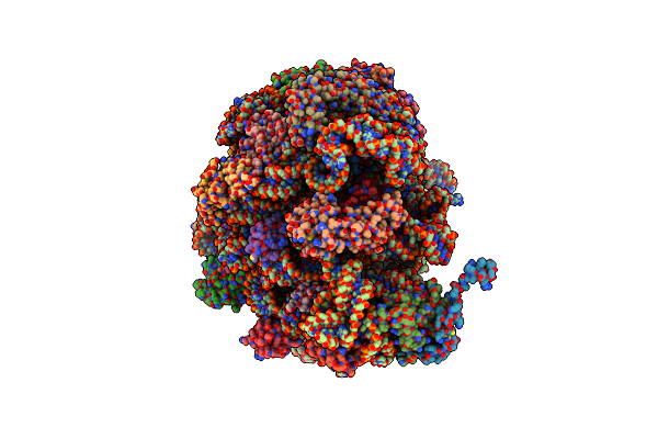
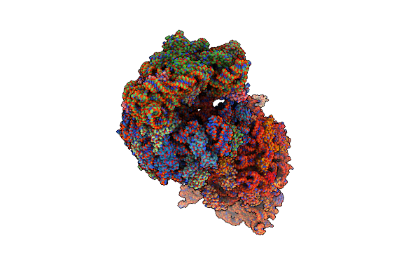
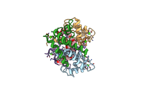
Cutibacterium Acnes 70S Ribosome With Mrna, P-Site Trna And Sarecycline Bound
Organism: Escherichia coli, Cutibacterium acnes
Method: ELECTRON MICROSCOPY Release Date: 2023-03-22 Classification: ANTIBIOTIC Ligands: V7A, MG, ZN |
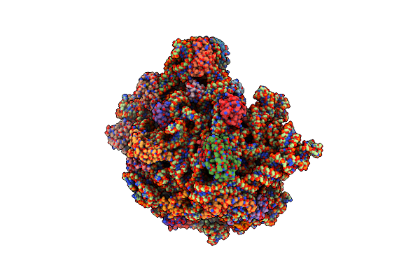 |
Cutibacterium Acnes 50S Ribosomal Subunit With P-Site Trna And Sarecycline Bound In The Local Refined Map
Organism: Cutibacterium acnes, Escherichia coli
Method: ELECTRON MICROSCOPY Release Date: 2023-03-15 Classification: ANTIBIOTIC Ligands: MG, ZN, V7A |
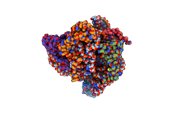 |
Cutibacterium Acnes 30S Ribosomal Subunit With Sarecycline Bound, Head Domain Only In The Local Refined Map
Organism: Cutibacterium acnes
Method: ELECTRON MICROSCOPY Release Date: 2023-03-15 Classification: ANTIBIOTIC Ligands: V7A, MG, ZN |
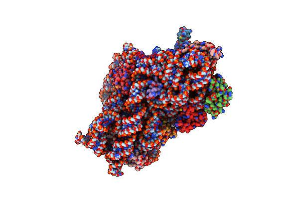 |
Cutibacterium Acnes 30S Ribosomal Subunit With Sarecycline Bound, Body Domain Only In The Local Refined Map
Organism: Escherichia coli, Cutibacterium acnes
Method: ELECTRON MICROSCOPY Release Date: 2023-03-15 Classification: ANTIBIOTIC Ligands: MG |
 |
Crystal Structure Of The Thermus Thermophilus 70S Ribosome In Complex With Sarecycline, Uuc-Mrna, And Deacylated P-Site Trna At 2.80A Resolution
Organism: Escherichia coli, Escherichia virus t4, Thermus thermophilus hb8
Method: X-RAY DIFFRACTION Resolution:2.80 Å Release Date: 2020-08-05 Classification: RIBOSOME Ligands: MG, ZN, V7A, SF4 |
 |
Crystal Structure Of The Thermus Thermophilus 70S Ribosome In Complex With Sarecycline, Uaa-Mrna, And Deacylated P-Site Trna At 3.00A Resolution
Organism: Escherichia coli, Escherichia virus t4, Thermus thermophilus hb8
Method: X-RAY DIFFRACTION Resolution:3.00 Å Release Date: 2020-08-05 Classification: RIBOSOME Ligands: MG, K, ZN, V7A, SF4 |
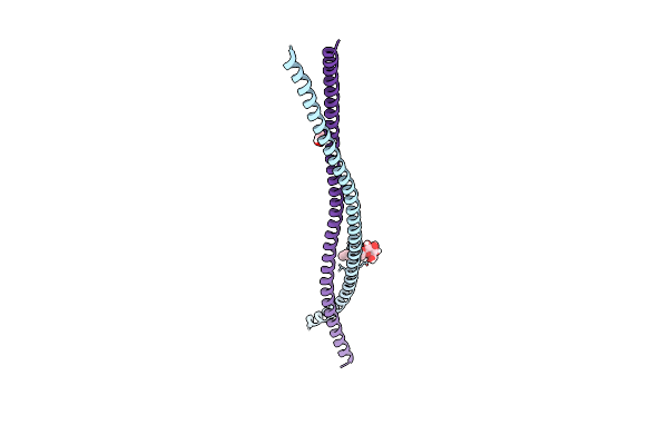 |
Crystal Structure Of The Heterocomplex Between Coil 2B Domains Of Wild-Type Keratin 1 (Krt1) And Keratin 10 (Krt10) Containing Mutation Cys401Ala
Organism: Homo sapiens
Method: X-RAY DIFFRACTION Resolution:2.07 Å Release Date: 2019-11-13 Classification: PROTEIN FIBRIL Ligands: BOG, GOL |
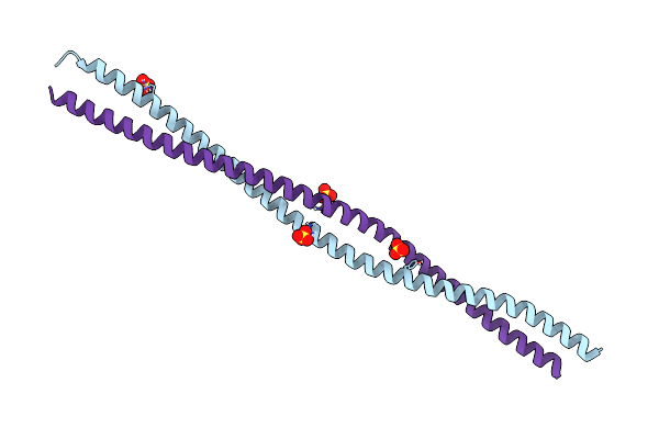 |
Crystal Structure Of The Heterocomplex Between Human Keratin 1 Coil 1B Containing S233L Mutation And Wild-Type Human Keratin 10 Coil 1B
Organism: Homo sapiens
Method: X-RAY DIFFRACTION Resolution:2.39 Å Release Date: 2019-05-15 Classification: PROTEIN FIBRIL Ligands: SO4 |
 |
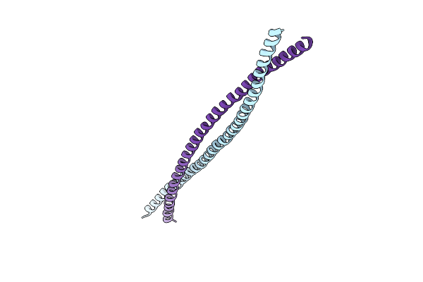
Crystal Structure Of The Wild-Type Heterocomplex Between Coil 1B Domains Of Human Intermediate Filament Proteins Keratin 1 (Krt1) And Keratin 10 (Krt10)
Organism: Homo sapiens
Method: X-RAY DIFFRACTION Resolution:2.98 Å Release Date: 2019-05-15 Classification: PROTEIN FIBRIL Ligands: CD, CO, NI |
 |
Crystal Structure Of The Heterocomplex Between Coil 2B Domains Of Human Intermediate Filament Proteins Keratin 1 (Krt1) And Keratin 10 (Krt10)
Organism: Homo sapiens
Method: X-RAY DIFFRACTION Resolution:3.30 Å Release Date: 2016-05-18 Classification: PROTEIN FIBRIL |
 |
Crystal Structure Of The N-Terminal Domain Of Human Profilaggrin At 2.2 A Resolution
Organism: Homo sapiens
Method: X-RAY DIFFRACTION Resolution:2.20 Å Release Date: 2015-04-01 Classification: METAL BINDING PROTEIN Ligands: CA, 2PE |
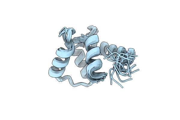 |
Solution Structure Of The Calcium-Loaded N-Terminal Sensor Domain Of Centrin
Organism: Chlamydomonas reinhardtii
Method: SOLUTION NMR Release Date: 2005-08-23 Classification: CELL CYCLE |
 |
X-Ray Structure Of Calcium-Loaded Calbindomodulin (A Calbindin D9K Re-Engineered To Undergo A Conformational Opening) At 1.44 A Resolution
Organism: Bos taurus
Method: X-RAY DIFFRACTION Resolution:1.44 Å Release Date: 2004-05-25 Classification: SIGNALING PROTEIN Ligands: CA, ZN |