
Search Count: 68
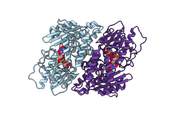 |
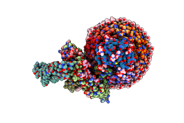
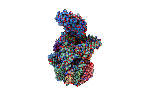
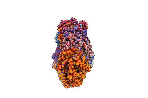
Organism: Mycolicibacterium smegmatis mc2 155
Method: ELECTRON MICROSCOPY Resolution:3.00 Å Release Date: 2025-01-22 Classification: MEMBRANE PROTEIN Ligands: FAD |
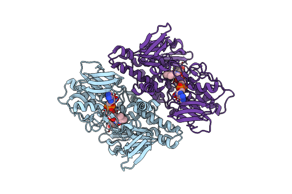 |
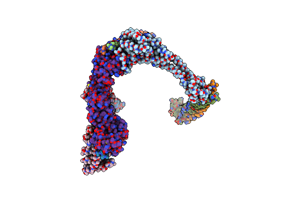
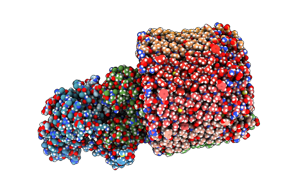
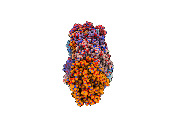
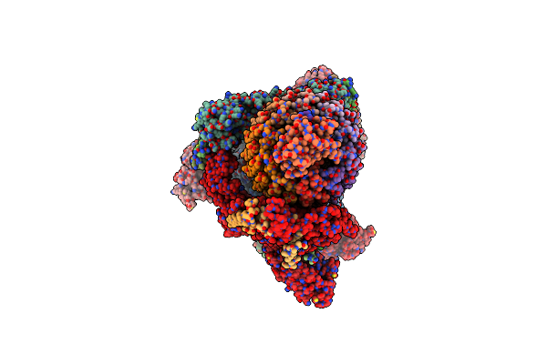
Structure Of Mycobacterial Ndh2 (Type Ii Nadh:Quinone Oxidoreductase) With 2-Mercapto-Quinazolinone Inhibitor.
Organism: Mycolicibacterium smegmatis mc2 155
Method: ELECTRON MICROSCOPY Resolution:3.00 Å Release Date: 2025-01-22 Classification: MEMBRANE PROTEIN Ligands: FAD, A1BNR |
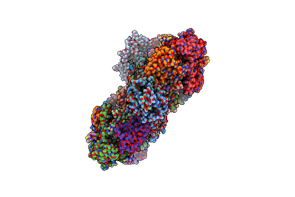 |
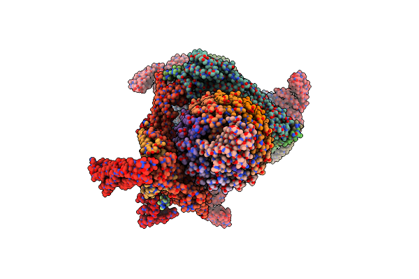
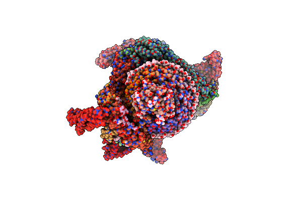
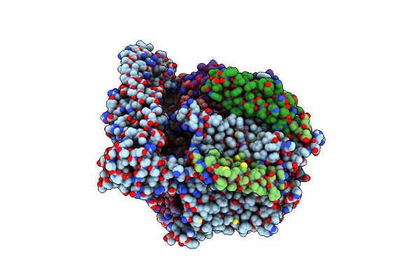
Organism: Mycolicibacterium smegmatis mc2 155
Method: ELECTRON MICROSCOPY Release Date: 2024-10-23 Classification: MEMBRANE PROTEIN Ligands: CU, HEA, CDL, HEM, MQ9, 9Y0, PLM, 9XX, HEC, 9YF, FES |
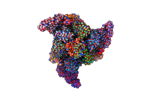 |
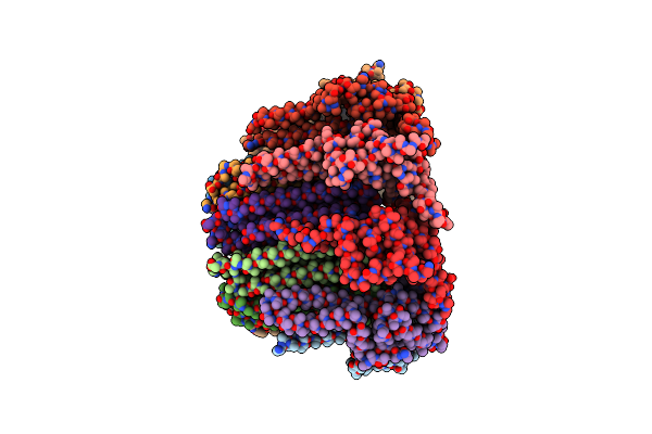
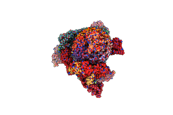
Organism: Legionella pneumophila subsp. pneumophila str. philadelphia 1, Rattus norvegicus
Method: ELECTRON MICROSCOPY Release Date: 2024-07-03 Classification: PROTON TRANSPORT Ligands: ADP |
 |
Organism: Rattus norvegicus
Method: ELECTRON MICROSCOPY Release Date: 2024-06-26 Classification: PROTON TRANSPORT Ligands: PC1, WJP, NAG, LP3, PTY, CLR |
 |
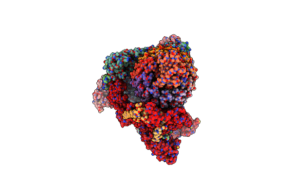
Synaptic Vesicle V-Atpase With Synaptophysin And Sidk, State 3, Peripheral Stalks
Organism: Rattus norvegicus
Method: ELECTRON MICROSCOPY Release Date: 2024-06-26 Classification: PROTON TRANSPORT |
 |
Organism: Legionella pneumophila subsp. pneumophila, Rattus norvegicus
Method: ELECTRON MICROSCOPY Release Date: 2024-06-26 Classification: PROTON TRANSPORT |
 |
Organism: Legionella pneumophila subsp. pneumophila, Rattus norvegicus
Method: ELECTRON MICROSCOPY Release Date: 2024-06-26 Classification: PROTON TRANSPORT |
 |
Organism: Legionella pneumophila subsp. pneumophila str. philadelphia 1, Rattus norvegicus
Method: ELECTRON MICROSCOPY Release Date: 2024-06-26 Classification: PROTON TRANSPORT Ligands: ADP, PC1, PTY, WJP, NAG, LP3, CLR |
 |
Organism: Saccharomyces cerevisiae
Method: ELECTRON MICROSCOPY Release Date: 2022-11-02 Classification: MEMBRANE PROTEIN |
 |
Organism: Saccharomyces cerevisiae
Method: ELECTRON MICROSCOPY Release Date: 2022-11-02 Classification: MEMBRANE PROTEIN |
 |
Organism: Saccharomyces cerevisiae
Method: ELECTRON MICROSCOPY Release Date: 2022-11-02 Classification: MEMBRANE PROTEIN |
 |
Organism: Saccharomyces cerevisiae
Method: ELECTRON MICROSCOPY Release Date: 2022-11-02 Classification: MEMBRANE PROTEIN |
 |
Organism: Mycolicibacterium smegmatis mc2 155
Method: ELECTRON MICROSCOPY Release Date: 2022-10-12 Classification: MEMBRANE PROTEIN Ligands: SF4, MQ9, FES, GTP, ZN, FMN, XP2 |
 |
Organism: Mycolicibacterium smegmatis mc2 155
Method: ELECTRON MICROSCOPY Release Date: 2022-10-12 Classification: MEMBRANE PROTEIN Ligands: SF4, MQ9, FES, GTP, FMN, ZN, XP2 |
 |
Organism: Mycolicibacterium smegmatis mc2 155
Method: ELECTRON MICROSCOPY Release Date: 2022-10-12 Classification: MEMBRANE PROTEIN Ligands: SF4, FES, GTP, FMN, ZN, MQ9, XP2 |
 |
Organism: Legionella pneumophila, Sus scrofa
Method: ELECTRON MICROSCOPY Release Date: 2022-07-06 Classification: MEMBRANE PROTEIN |
 |
Organism: Legionella pneumophila, Sus scrofa
Method: ELECTRON MICROSCOPY Release Date: 2022-07-06 Classification: MEMBRANE PROTEIN Ligands: ADP |
 |
Organism: Legionella pneumophila, Sus scrofa
Method: ELECTRON MICROSCOPY Release Date: 2022-07-06 Classification: MEMBRANE PROTEIN Ligands: ADP |
 |
Organism: Legionella pneumophila, Sus scrofa
Method: ELECTRON MICROSCOPY Release Date: 2022-07-06 Classification: MEMBRANE PROTEIN Ligands: ADP |