
Search Count: 324
 |
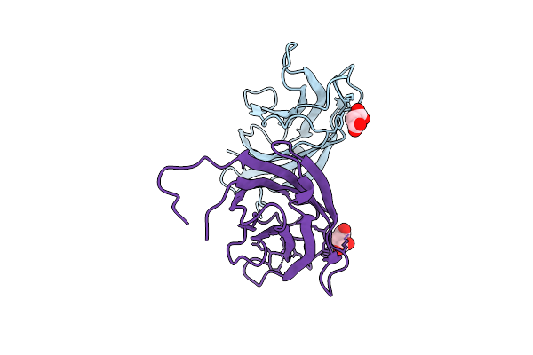
Crystal Structure Of A Calcium Bound C2 Domain Containing Protein From Trichomonas Vaginalis
Organism: Trichomonas vaginalis g3
Method: X-RAY DIFFRACTION Release Date: 2025-12-17 Classification: LIPID BINDING PROTEIN Ligands: 1PE, CL, PEG, PGE |
 |
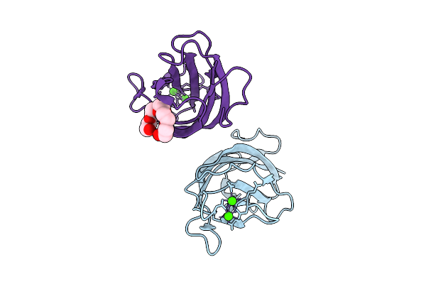
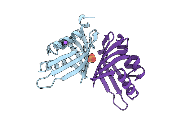
Crystal Structure Of A Calcium Bound C2 Domain Containing Protein From Trichomonas Vaginalis (P21 Form)
Organism: Trichomonas vaginalis g3
Method: X-RAY DIFFRACTION Release Date: 2025-12-17 Classification: LIPID BINDING PROTEIN Ligands: PG4, CA, MES, 1PE |
 |
Crystal Structure Of A Calcium Bound C2 Domain Containing Protein From Trichomonas Vaginalis (Orthorhombic P Form)
Organism: Trichomonas vaginalis g3
Method: X-RAY DIFFRACTION Release Date: 2025-12-17 Classification: LIPID BINDING PROTEIN Ligands: CA, 1PE |
 |
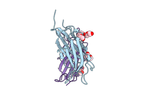
Crystal Structure Of A C2 Domain Containing Protein From Trichomonas Vaginalis
Organism: Trichomonas vaginalis g3
Method: X-RAY DIFFRACTION Release Date: 2025-12-17 Classification: LIPID BINDING PROTEIN Ligands: GOL, PGE |
 |
Crystal Structure Of A Malonate Bound C2 Domain Containing Protein From Trichomonas Vaginalis (P21 Form)
Organism: Trichomonas vaginalis g3
Method: X-RAY DIFFRACTION Release Date: 2025-12-17 Classification: LIPID BINDING PROTEIN Ligands: MLI, CL, NA |
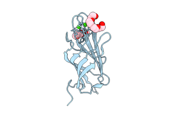 |
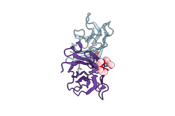
Crystal Structure Of A Calcium Bound C2 Domain Containing Protein From Trichomonas Vaginalis (P61 Form)
Organism: Trichomonas vaginalis g3
Method: X-RAY DIFFRACTION Release Date: 2025-12-17 Classification: LIPID BINDING PROTEIN Ligands: CA, CL, PG4 |
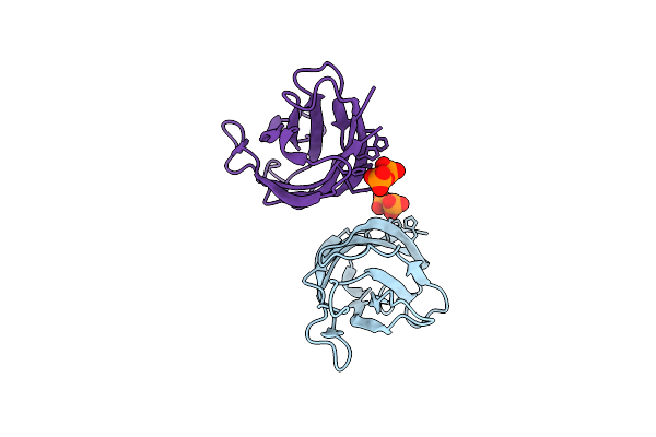 |
Crystal Structure Of A C2 Domain Containing Protein From Trichomonas Vaginalis In Complex With Pyrophosphate
Organism: Trichomonas vaginalis g3
Method: X-RAY DIFFRACTION Release Date: 2025-12-17 Classification: LIPID BINDING PROTEIN Ligands: PPV |
 |
Crystal Structure Of A Phage Catechol 1,2-Dioxygenase Identified From A Soil Metagenomic Survey
Organism: Metagenome
Method: X-RAY DIFFRACTION Release Date: 2025-10-08 Classification: OXIDOREDUCTASE Ligands: FE |
 |
Crystal Structure Of A Snoal-Like Domain-Containing Protein From Mycobacterium Ulcerans (Orthorhombic C Form)
Organism: Mycobacterium ulcerans agy99
Method: X-RAY DIFFRACTION Release Date: 2025-09-24 Classification: ISOMERASE Ligands: PG4 |
 |
Crystal Structure Of A Snoal-Like Domain-Containing Protein From Mycobacterium Ulcerans
Organism: Mycobacterium ulcerans agy99
Method: X-RAY DIFFRACTION Release Date: 2025-07-23 Classification: ISOMERASE Ligands: PO4, NA |
 |
Organism: Xanthomonas sacchari
Method: X-RAY DIFFRACTION Resolution:2.30 Å Release Date: 2025-05-21 Classification: SUGAR BINDING PROTEIN Ligands: EDO, A1BNY |
 |
Organism: Streptomyces sp.
Method: X-RAY DIFFRACTION Resolution:1.45 Å Release Date: 2025-05-21 Classification: SUGAR BINDING PROTEIN Ligands: BTB, EDO |
 |
Crystal Structure Of A Putative Phage Endolysin Identified From A Metagenomic Survey Of Prosser, Washington Soil (Pwe1)
Organism: Metagenome
Method: X-RAY DIFFRACTION Resolution:2.15 Å Release Date: 2025-05-21 Classification: HYDROLASE Ligands: SO4, BR |
 |
Crystal Structure Of A Putative Phage Endolysin Identified From A Metagenomic Survey Of Prosser, Washington Soil (Pwe2)
Organism: Metagenome
Method: X-RAY DIFFRACTION Resolution:1.50 Å Release Date: 2025-05-21 Classification: HYDROLASE Ligands: MG, EDO, CL |
 |
Solution Structure Of An Uncharacterized Protein Containing A Duf1893 Domain From Borrelia Burgdorferi, A Pathogen Responsible For Lyme Disease. Seattle Structural Genomics Center For Infectious Disease Target Bobua.17726.A.A1.
Organism: Borreliella burgdorferi b31
Method: SOLUTION NMR Release Date: 2024-12-04 Classification: UNKNOWN FUNCTION |
 |
Solution Structure Of The Translation Initiation Factor If-1 From Neisseria Gonorrhoeae (Nccp11945). Seattle Structural Genomics Center For Infectious Disease Target Negoa.17902.A
Organism: Neisseria gonorrhoeae (strain atcc 700825 / fa 1090)
Method: SOLUTION NMR Release Date: 2024-10-16 Classification: RNA BINDING PROTEIN |
 |
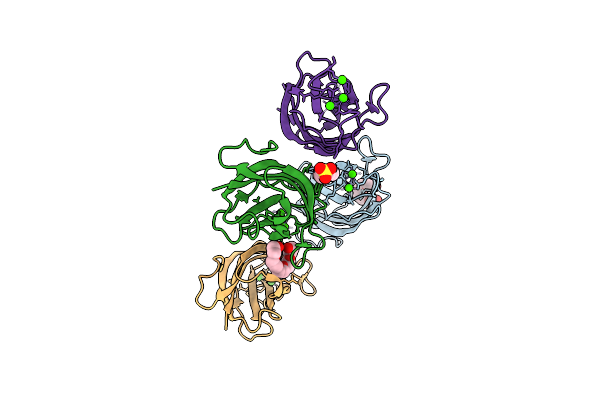
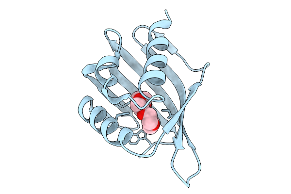
Crystal Structure Of Adp-Ribose Diphosphatase From Klebsiella Pneumoniae (Atp Bound)
Organism: Klebsiella pneumoniae
Method: X-RAY DIFFRACTION Resolution:1.55 Å Release Date: 2024-10-16 Classification: HYDROLASE Ligands: ATP |
 |
Crystal Structure Of Adp-Ribose Diphosphatase From Klebsiella Pneumoniae (Adp/Mg Bound)
Organism: Klebsiella pneumoniae
Method: X-RAY DIFFRACTION Resolution:1.35 Å Release Date: 2024-10-16 Classification: HYDROLASE Ligands: MG, ADP |
 |
Crystal Structure Of Adp-Ribose Diphosphatase From Klebsiella Pneumoniae (Gdp/Mg Bound)
Organism: Klebsiella pneumoniae
Method: X-RAY DIFFRACTION Resolution:1.35 Å Release Date: 2024-10-16 Classification: HYDROLASE Ligands: GDP, MG |
 |
Crystal Structure Of Adp-Ribose Diphosphatase From Klebsiella Pneumoniae (Gmp Bound)
Organism: Klebsiella pneumoniae
Method: X-RAY DIFFRACTION Resolution:1.40 Å Release Date: 2024-10-16 Classification: HYDROLASE Ligands: 5GP, MG |