
Search Count: 34
 |
Organism: Homo sapiens
Method: ELECTRON MICROSCOPY Release Date: 2025-05-14 Classification: MEMBRANE PROTEIN |
 |
Organism: Drosophila melanogaster
Method: ELECTRON MICROSCOPY Release Date: 2025-01-29 Classification: SIGNALING PROTEIN Ligands: NAG |
 |
Organism: Drosophila melanogaster
Method: ELECTRON MICROSCOPY Release Date: 2025-01-29 Classification: SIGNALING PROTEIN Ligands: NAG |
 |
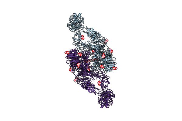
Cryo-Em Structure Of Bride Of Sevenless Extracellular Domain (Dimer, Sevenless-Bound Form)
Organism: Drosophila melanogaster
Method: ELECTRON MICROSCOPY Release Date: 2025-01-29 Classification: SIGNALING PROTEIN Ligands: NAG |
 |
Organism: Drosophila melanogaster
Method: ELECTRON MICROSCOPY Release Date: 2025-01-29 Classification: SIGNALING PROTEIN Ligands: NAG |
 |
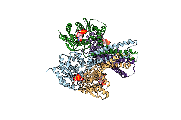
Cryo-Em Structure Of Sevenless Extracellular Domain (Composite Map Of The Dimer, Ph 4.6)
Organism: Drosophila melanogaster
Method: ELECTRON MICROSCOPY Release Date: 2025-01-29 Classification: SIGNALING PROTEIN Ligands: NAG |
 |
Organism: Colwellia psychrerythraea
Method: X-RAY DIFFRACTION Resolution:2.40 Å Release Date: 2017-08-09 Classification: MEMBRANE PROTEIN Ligands: CD |
 |
Organism: Sulfolobus acidocaldarius
Method: X-RAY DIFFRACTION Resolution:1.60 Å Release Date: 2017-07-12 Classification: MEMBRANE PROTEIN |
 |
Organism: Sulfolobus acidocaldarius
Method: X-RAY DIFFRACTION Resolution:1.90 Å Release Date: 2017-06-21 Classification: MEMBRANE PROTEIN Ligands: MG |
 |
Organism: Sulfolobus acidocaldarius
Method: X-RAY DIFFRACTION Resolution:2.40 Å Release Date: 2017-06-21 Classification: MEMBRANE PROTEIN Ligands: SO4 |
 |
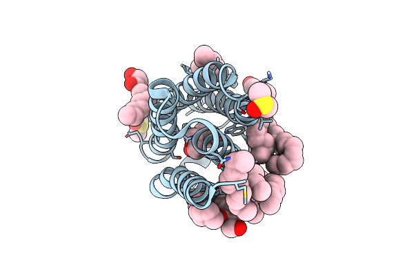
Structure Of The Polyisoprenyl-Phosphate Glycosyltransferase Gtrb (F215A Mutant)
Organism: Synechocystis sp. (strain pcc 6803 / kazusa)
Method: X-RAY DIFFRACTION Resolution:3.00 Å Release Date: 2016-01-06 Classification: TRANSFERASE Ligands: UDP, MG |
 |
Organism: Synechocystis sp. (strain pcc 6803 / kazusa)
Method: X-RAY DIFFRACTION Resolution:3.19 Å Release Date: 2016-01-06 Classification: TRANSFERASE Ligands: UDP, MG |
 |
Organism: Bacillus cereus
Method: X-RAY DIFFRACTION Resolution:1.60 Å Release Date: 2015-04-22 Classification: MEMBRANE PROTEIN Ligands: MPG, DMS |
 |
Organism: Bacillus cereus
Method: X-RAY DIFFRACTION Resolution:4.10 Å Release Date: 2015-02-11 Classification: MEMBRANE PROTEIN |
 |
Organism: Bacillus cereus
Method: X-RAY DIFFRACTION Resolution:2.01 Å Release Date: 2015-02-11 Classification: MEMBRANE PROTEIN Ligands: MPG, LMU, CAC, PGE |
 |
Organism: Bacillus cereus
Method: X-RAY DIFFRACTION Resolution:3.49 Å Release Date: 2015-01-28 Classification: MEMBRANE PROTEIN Ligands: PKA |
 |
Organism: Bacillus cereus
Method: X-RAY DIFFRACTION Resolution:1.70 Å Release Date: 2015-01-28 Classification: MEMBRANE PROTEIN Ligands: MPG |
 |
Organism: Bacillus cereus
Method: X-RAY DIFFRACTION Resolution:1.70 Å Release Date: 2015-01-28 Classification: MEMBRANE PROTEIN Ligands: MPG, DMS |
 |
Crystal Structure Of A Bacterial Bestrophin Homolog From Klebsiella Pneumoniae By Zn-Sad Phasing
Organism: Klebsiella pneumoniae uhkpc96
Method: X-RAY DIFFRACTION Resolution:2.90 Å Release Date: 2014-10-01 Classification: MEMBRANE PROTEIN Ligands: ZN |
 |
Crystal Structure Of A Bacterial Bestrophin Homolog From Klebsiella Pneumoniae
Organism: Klebsiella pneumoniae uhkpc96
Method: X-RAY DIFFRACTION Resolution:2.30 Å Release Date: 2014-10-01 Classification: MEMBRANE PROTEIN Ligands: ZN |