
Search Count: 8
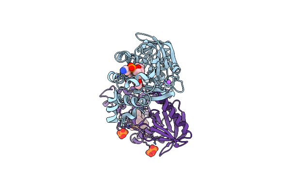 |
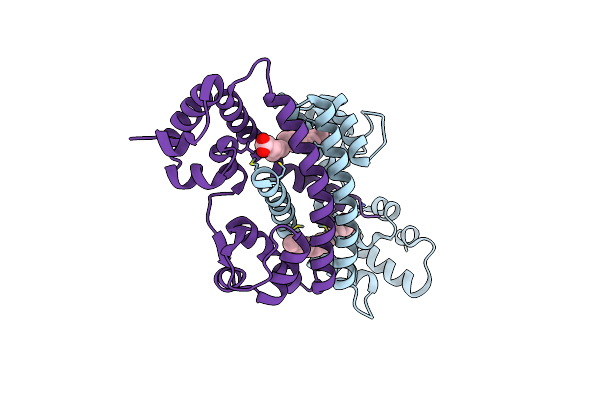
Crystal Structure Of The Neurofibromin Sec14-Ph Module Containing The Patient Derived Mutation I1584V
Organism: Homo sapiens
Method: X-RAY DIFFRACTION Resolution:2.65 Å Release Date: 2010-12-08 Classification: LIPID BINDING PROTEIN Ligands: PEV, NA, POP |
 |
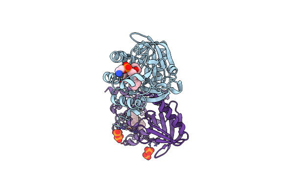
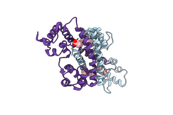
Crystal Structure Of Neurofibromins Sec14-Ph Module Containing A Patient Derived Duplication (Td)
Organism: Homo sapiens
Method: X-RAY DIFFRACTION Resolution:2.52 Å Release Date: 2010-12-08 Classification: LIPID BINDING PROTEIN Ligands: SO4, MG, PEV |
 |
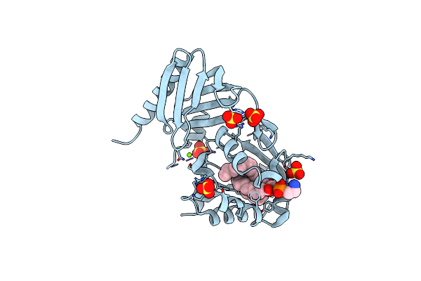
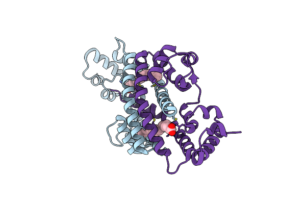
Organism: Homo sapiens
Method: X-RAY DIFFRACTION Resolution:2.19 Å Release Date: 2010-12-08 Classification: LIPID BINDING PROTEIN Ligands: PTY, POP |
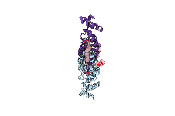 |
Structure Of Rv1264N, The Regulatory Domain Of The Mycobacterial Adenylyl Cylcase Rv1264, At Ph 6.0
Organism: Mycobacterium tuberculosis
Method: X-RAY DIFFRACTION Resolution:1.60 Å Release Date: 2006-11-07 Classification: LYASE Ligands: OLA, 1PE |
 |
Structure Of Rv1264N, The Regulatory Domain Of The Mycobacterial Adenylyl Cylcase Rv1264, At Ph 8.5
Organism: Mycobacterium tuberculosis
Method: X-RAY DIFFRACTION Resolution:2.35 Å Release Date: 2006-11-07 Classification: LYASE Ligands: OLA |
 |
Structure Of Rv1264N, The Regulatory Domain Of The Mycobacterial Adenylyl Cylcase Rv1264, At Ph 5.3
Organism: Mycobacterium tuberculosis
Method: X-RAY DIFFRACTION Resolution:2.68 Å Release Date: 2006-11-07 Classification: LYASE Ligands: OLA |
 |
Structure Of Rv1264N, The Regulatory Domain Of The Mycobacterial Adenylyl Cylcase Rv1264, With A Salt Precipitant
Organism: Mycobacterium tuberculosis
Method: X-RAY DIFFRACTION Resolution:2.28 Å Release Date: 2006-11-07 Classification: LYASE Ligands: CL, OLA |
 |