
Search Count: 28
 |
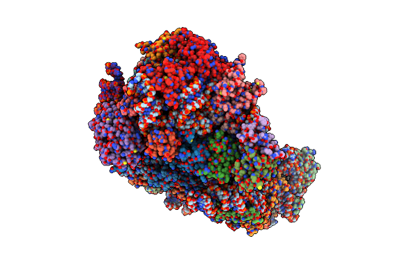
Time-Resolved Cryo-Em Study Of The 70S Recycling By The Hflx:Control-Apo-70S At 900Ms
Organism: Escherichia coli
Method: ELECTRON MICROSCOPY Release Date: 2023-12-06 Classification: RIBOSOME |
 |
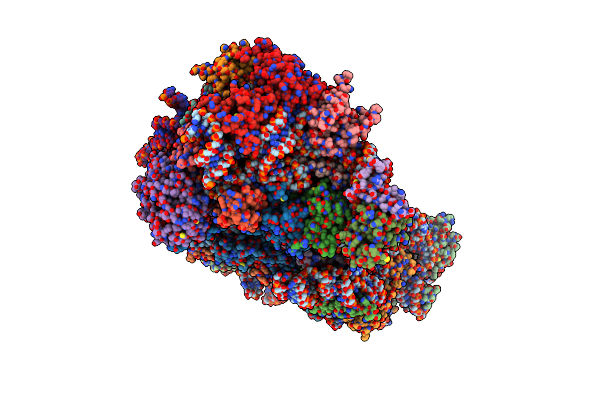
Time-Resolved Cryo-Em Study Of The 70S Recycling By The Hflx:2Nd Intermediate
Organism: Escherichia coli
Method: ELECTRON MICROSCOPY Release Date: 2023-12-06 Classification: RIBOSOME |
 |
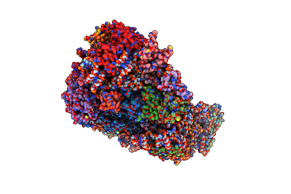
Time-Resolved Cryo-Em Study Of The 70S Recycling By The Hflx:1St Intermediate
Organism: Escherichia coli
Method: ELECTRON MICROSCOPY Release Date: 2023-12-06 Classification: RIBOSOME |
 |
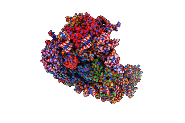
Time-Resolved Cryo-Em Study Of The 70S Recycling By The Hflx:3Rd Intermediate
Organism: Escherichia coli
Method: ELECTRON MICROSCOPY Release Date: 2023-12-06 Classification: RIBOSOME Ligands: GTP |
 |
Structure Of The Hcv Ires Bound To The 40S Ribosomal Subunit, Head Open. Structure 9(Delta Dii)
Organism: Hepatitis c virus (isolate 1), Oryctolagus cuniculus
Method: ELECTRON MICROSCOPY Release Date: 2022-07-27 Classification: RIBOSOME Ligands: ZN |
 |
Structure Of The Wt Ires And 40S Ribosome Binary Complex, Open Conformation. Structure 10(Wt)
Organism: Hepatitis c virus (isolate 1), Oryctolagus cuniculus
Method: ELECTRON MICROSCOPY Release Date: 2022-07-27 Classification: RIBOSOME Ligands: ZN |
 |
Structure Of The Wt Ires And 40S Ribosome Ternary Complex, Open Conformation. Structure 11(Wt)
Organism: Homo sapiens, Hepatitis c virus (isolate 1), Oryctolagus cuniculus
Method: ELECTRON MICROSCOPY Release Date: 2022-07-27 Classification: RIBOSOME Ligands: ZN |
 |
Structure Of The Wt Ires Eif2-Containing 48S Initiation Complex, Closed Conformation. Structure 12(Wt).
Organism: Homo sapiens, Hepatitis c virus (isolate 1), Oryctolagus cuniculus
Method: ELECTRON MICROSCOPY Release Date: 2022-07-27 Classification: RIBOSOME Ligands: ZN |
 |
Structure Of The Delta Dii Ires Eif2-Containing 48S Initiation Complex, Closed Conformation. Structure 12(Delta Dii).
Organism: Homo sapiens, Hepatitis c virus (isolate 1), Oryctolagus cuniculus
Method: ELECTRON MICROSCOPY Release Date: 2022-07-27 Classification: RIBOSOME Ligands: ZN |
 |
Structure Of The Wt Ires Eif5B-Containing Pre-48S Initiation Complex, Open Conformation. Structure 14(Wt)
Organism: Homo sapiens, Hepatitis c virus genotype 1a, Oryctolagus cuniculus
Method: ELECTRON MICROSCOPY Release Date: 2022-07-20 Classification: RIBOSOME Ligands: ZN, GTP, MG, NA |
 |
Structure Of The Hcv Ires Binding To The 40S Ribosomal Subunit, Closed Conformation. Structure 1(Delta Dii)
Organism: Hepatitis c virus (isolate 1), Oryctolagus cuniculus
Method: ELECTRON MICROSCOPY Release Date: 2022-07-13 Classification: RIBOSOME Ligands: ZN |
 |
Structure Of The Hcv Ires Binding To The 40S Ribosomal Subunit, Closed Conformation. Structure 2(Delta Dii)
Organism: Hepatitis c virus (isolate 1), Oryctolagus cuniculus
Method: ELECTRON MICROSCOPY Release Date: 2022-07-13 Classification: RIBOSOME Ligands: ZN |
 |
Structure Of The Hcv Ires Binding To The 40S Ribosomal Subunit, Closed Conformation. Structure 3(Delta Dii)
Organism: Hepacivirus c, Oryctolagus cuniculus
Method: ELECTRON MICROSCOPY Release Date: 2022-07-13 Classification: RIBOSOME Ligands: ZN |
 |
Structure Of The Hcv Ires Binding To The 40S Ribosomal Subunit, Closed Conformation. Structure 4(Delta Dii)
Organism: Hepatitis c virus genotype 1a, Oryctolagus cuniculus
Method: ELECTRON MICROSCOPY Release Date: 2022-07-13 Classification: RIBOSOME Ligands: ZN |
 |
Structure Of The Hcv Ires Binding To The 40S Ribosomal Subunit, Closed Conformation. Structure 5(Delta Dii)
Organism: Hepatitis c virus genotype 1a, Oryctolagus cuniculus
Method: ELECTRON MICROSCOPY Release Date: 2022-07-13 Classification: RIBOSOME Ligands: ZN |
 |
Structure Of The Hcv Ires Bound To The 40S Ribosomal Subunit, Closed Conformation. Structure 6(Delta Dii)
Organism: Hepatitis c virus genotype 1a, Oryctolagus cuniculus
Method: ELECTRON MICROSCOPY Release Date: 2022-07-13 Classification: RIBOSOME Ligands: ZN |
 |
Structure Of The Hcv Ires Bound To The 40S Ribosomal Subunit, Head Opening. Structure 7(Delta Dii)
Organism: Hepatitis c virus (isolate 1), Oryctolagus cuniculus
Method: ELECTRON MICROSCOPY Release Date: 2022-07-13 Classification: RIBOSOME Ligands: ZN |
 |
Structure Of The Hcv Ires Bound To The 40S Ribosomal Subunit, Head Opening. Structure 8(Delta Dii)
Organism: Hepatitis c virus (isolate 1), Oryctolagus cuniculus
Method: ELECTRON MICROSCOPY Release Date: 2022-07-13 Classification: RIBOSOME Ligands: ZN |
 |
Structure Of The Wt Ires W/O Eif2 48S Initiation Complex, Closed Conformation. Structure 13(Wt)
Organism: Hepatitis c virus (isolate 1), Oryctolagus cuniculus
Method: ELECTRON MICROSCOPY Release Date: 2022-07-13 Classification: RIBOSOME Ligands: ZN |
 |
Structure Of The Delta Dii Ires W/O Eif2 48S Initiation Complex, Closed Conformation. Structure 13(Delta Dii)
Organism: Hepatitis c virus (isolate 1), Oryctolagus cuniculus
Method: ELECTRON MICROSCOPY Release Date: 2022-07-13 Classification: RIBOSOME Ligands: ZN |