
Search Count: 31
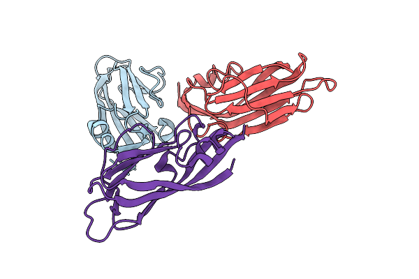 |
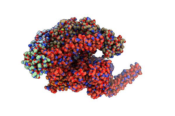
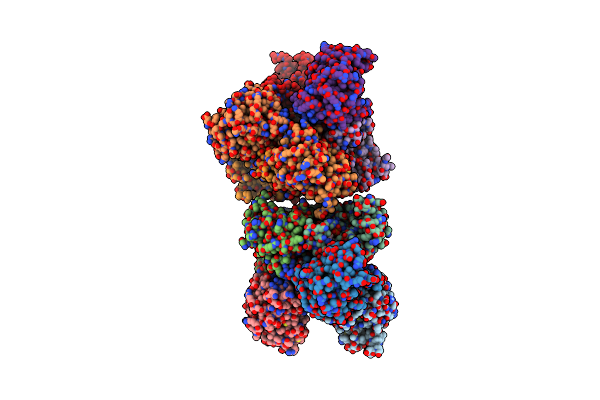
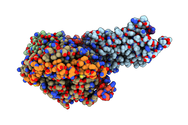
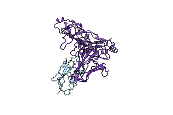
Cryoem Structure Of Modified Turnip Yellows Virus Devoid Of Minor Capsid Protein Readthrough Domain
Organism: Turnip yellows virus
Method: ELECTRON MICROSCOPY Release Date: 2025-05-07 Classification: VIRUS |
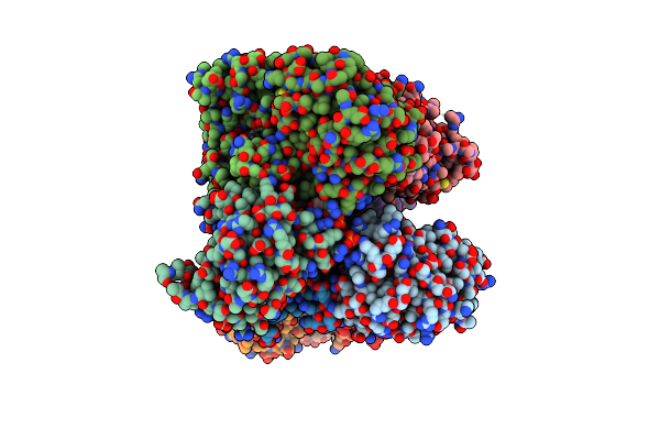
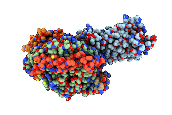
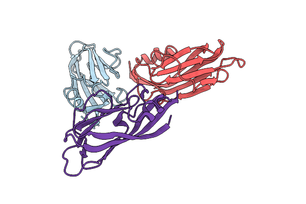 |
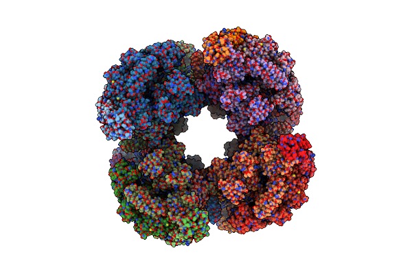
Organism: Turnip yellows virus
Method: ELECTRON MICROSCOPY Release Date: 2024-06-05 Classification: VIRUS |
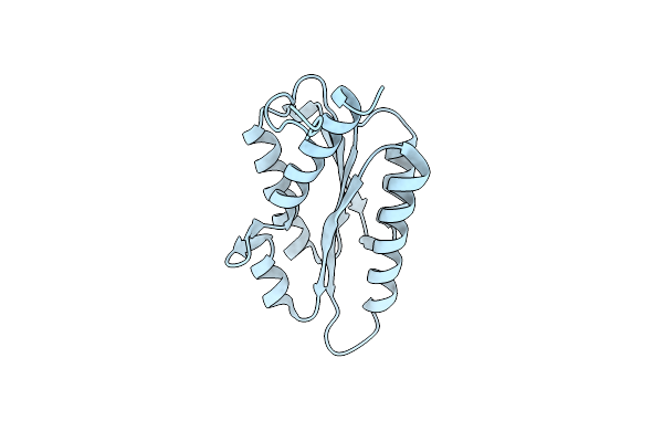 |
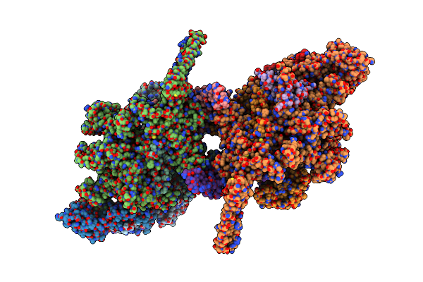
Organism: Vicia faba
Method: ELECTRON MICROSCOPY Release Date: 2024-05-22 Classification: TRANSFERASE |
 |
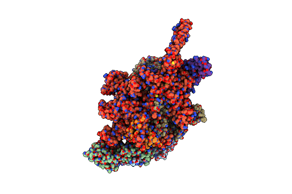
Organism: Mycobacterium tuberculosis h37rv
Method: ELECTRON MICROSCOPY Release Date: 2023-02-08 Classification: TRANSCRIPTION Ligands: ZN, MG |
 |
Protomeric Substructure From An Octameric Assembly Of M. Tuberculosis Rna Polymerase In Complex With Sigma-B Initiation Factor
Organism: Mycobacterium tuberculosis
Method: ELECTRON MICROSCOPY Release Date: 2023-02-08 Classification: TRANSCRIPTION Ligands: ZN, MG |
 |
Cryo-Em Structure Of Mycobacterium Tuberculosis Rna Polymerase Holoenzyme Octamer Comprising Sigma Factor Sigb
Organism: Mycobacterium tuberculosis (strain atcc 25618 / h37rv)
Method: ELECTRON MICROSCOPY Release Date: 2022-11-16 Classification: TRANSCRIPTION Ligands: ZN, MG |
 |
Cryo-Em Structure Of Mycobacterium Tuberculosis Rna Polymerase Holoenzyme Dimer Comprising Sigma Factor Sigb
Organism: Mycobacterium tuberculosis h37rv
Method: ELECTRON MICROSCOPY Release Date: 2022-11-16 Classification: TRANSCRIPTION Ligands: ZN, MG |
 |
Cryo-Em Structure Of Mycobacterium Tuberculosis Rna Polymerase Holoenzyme Comprising Sigma Factor Sigb
Organism: Mycobacterium tuberculosis (strain atcc 25618 / h37rv)
Method: ELECTRON MICROSCOPY Resolution:3.84 Å Release Date: 2022-09-21 Classification: TRANSCRIPTION Ligands: ZN, MG |
 |
Organism: Homo sapiens, Synthetic construct
Method: ELECTRON MICROSCOPY Release Date: 2022-09-14 Classification: MEMBRANE PROTEIN |
 |
Organism: Homo sapiens, Synthetic construct
Method: ELECTRON MICROSCOPY Release Date: 2022-09-14 Classification: MEMBRANE PROTEIN |
 |
Crystal Structure Of Umpk From M. Tuberculosis In Complex With Udp And Utp (C2 Form)
Organism: Mycobacterium tuberculosis h37rv
Method: X-RAY DIFFRACTION Resolution:3.12 Å Release Date: 2022-03-02 Classification: TRANSFERASE Ligands: UTP, UDP |
 |
Crystal Structure Of Umpk From M. Tuberculosis In Complex With Udp And Utp (P21212 Form)
Organism: Mycobacterium tuberculosis h37rv
Method: X-RAY DIFFRACTION Resolution:3.33 Å Release Date: 2022-03-02 Classification: TRANSFERASE Ligands: UDP, UTP |
 |
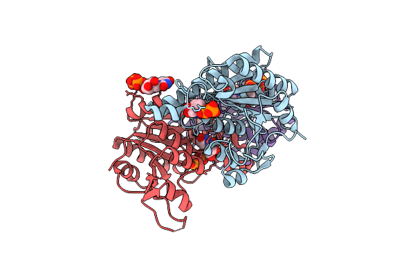
Cryoem Structure Of The Transcription Termination Factor Rho From Mycobacterium Tuberculosis
Organism: Mycobacterium tuberculosis
Method: ELECTRON MICROSCOPY Release Date: 2022-02-09 Classification: TRANSCRIPTION Ligands: ATP, MG |
 |
Cryoem Structure Of Mycobacterium Tuberculosis Ump Kinase (Umpk) In Complex With Udp And Utp
Organism: Mycobacterium tuberculosis
Method: ELECTRON MICROSCOPY Release Date: 2022-01-12 Classification: TRANSFERASE Ligands: UDP, UTP |
 |
Organism: Homo sapiens, Lama glama
Method: ELECTRON MICROSCOPY Release Date: 2021-06-02 Classification: MEMBRANE PROTEIN |
 |
Organism: Homo sapiens, Lama glama
Method: ELECTRON MICROSCOPY Release Date: 2021-06-02 Classification: MEMBRANE PROTEIN |
 |
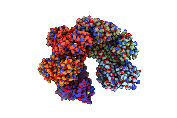
Organism: Faba bean necrotic stunt virus
Method: ELECTRON MICROSCOPY Release Date: 2020-07-15 Classification: VIRUS |
 |
Organism: Broad bean stain virus
Method: ELECTRON MICROSCOPY Release Date: 2019-05-01 Classification: VIRUS |
 |
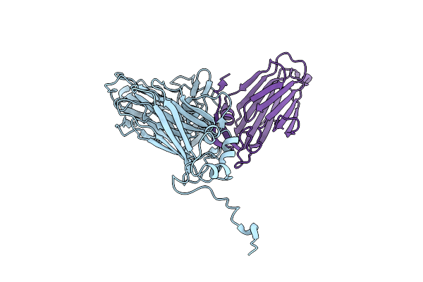
Cryo Electron Microscopy Structure Of Grapevine Fanleaf Virus Complex With Nanobody
Organism: Camelus dromedarius, Grapevine fanleaf virus
Method: ELECTRON MICROSCOPY Resolution:2.80 Å Release Date: 2016-01-20 Classification: VIRUS |
 |
Organism: Lactococcus phage p2
Method: X-RAY DIFFRACTION Resolution:5.46 Å Release Date: 2014-07-09 Classification: VIRAL PROTEIN Ligands: CA |