Search Count: 32
 |
Organism: Bdellovibrio bacteriovorus hd100
Method: X-RAY DIFFRACTION Release Date: 2025-10-22 Classification: SIGNALING PROTEIN |
 |
Cryo-Em Structure Of The Proximal End Of Bacteriophage T5 Tail : P142 Tail Terminator Protein Hexamer And Pb6 Tail Tube Protein Trimer
Organism: Escherichia phage t5
Method: ELECTRON MICROSCOPY Release Date: 2023-11-01 Classification: VIRAL PROTEIN |
 |
Cryo-Em Structure Of The Proximal End Of Bacteriophage T5 Tail, After Interaction With Its Receptor : P142 Tail Terminator Protein Hexamer And Pb6 Tail Tube Protein Trimer
Organism: Escherichia phage t5
Method: ELECTRON MICROSCOPY Release Date: 2023-11-01 Classification: VIRAL PROTEIN |
 |
Organism: Bdellovibrio bacteriovorus hd100
Method: X-RAY DIFFRACTION Resolution:2.17 Å Release Date: 2023-10-25 Classification: UNKNOWN FUNCTION Ligands: GOL, EDO |
 |
Crystal Structure Of The N-Terminal Domain Of The Cryptic Surface Protein (Cd630_25440) From Clostridium Difficile.
Organism: Clostridioides difficile 630
Method: X-RAY DIFFRACTION Resolution:2.00 Å Release Date: 2023-05-10 Classification: UNKNOWN FUNCTION Ligands: CL, EDO, GOL |
 |
Organism: Escherichia phage t5
Method: ELECTRON MICROSCOPY Release Date: 2023-02-08 Classification: VIRAL PROTEIN |
 |
Organism: Escherichia phage t5
Method: ELECTRON MICROSCOPY Release Date: 2023-02-08 Classification: VIRUS LIKE PARTICLE |
 |
Tail Tip Of Siphophage T5 : Full Complex After Interaction With Its Bacterial Receptor Fhua
Organism: Escherichia phage t5
Method: ELECTRON MICROSCOPY Release Date: 2023-02-08 Classification: VIRAL PROTEIN |
 |
Tail Tip Of Siphophage T5 : Bent Fibre After Interaction With Its Bacterial Receptor Fhua
Organism: Escherichia phage t5
Method: ELECTRON MICROSCOPY Release Date: 2023-02-08 Classification: VIRAL PROTEIN |
 |
Organism: Escherichia phage t5
Method: ELECTRON MICROSCOPY Release Date: 2023-02-08 Classification: VIRAL PROTEIN |
 |
Tail Tip Of Siphophage T5 : Open Cone After Interaction With Bacterial Receptor Fhua
Organism: Escherichia phage t5
Method: ELECTRON MICROSCOPY Release Date: 2023-02-08 Classification: VIRAL PROTEIN |
 |
Organism: Escherichia coli, Escherichia phage t5
Method: ELECTRON MICROSCOPY Release Date: 2023-02-08 Classification: VIRAL PROTEIN Ligands: DDQ, LU9 |
 |
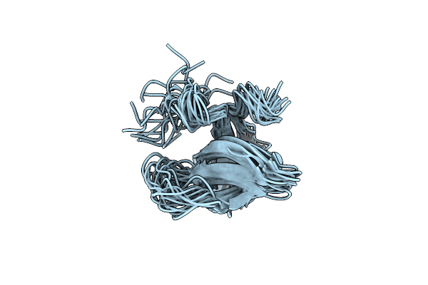
Organism: Escherichia phage t5
Method: SOLUTION NMR Release Date: 2022-12-28 Classification: VIRAL PROTEIN |
 |
Organism: Escherichia phage t5
Method: ELECTRON MICROSCOPY Release Date: 2022-12-21 Classification: VIRAL PROTEIN |
 |
Organism: Bdellovibrio bacteriovorus (strain atcc 15356 / dsm 50701 / ncib 9529 / hd100)
Method: X-RAY DIFFRACTION Resolution:1.50 Å Release Date: 2021-05-05 Classification: UNKNOWN FUNCTION Ligands: MPD, GOL, TBU |
 |
Organism: Caldicellulosiruptor bescii (strain atcc baa-1888 / dsm 6725 / z-1320)
Method: X-RAY DIFFRACTION Resolution:1.90 Å Release Date: 2021-04-14 Classification: TRANSFERASE Ligands: SAM, SAH, EDO |
 |
Crystal Structure Of The Protease 1 (E29A,E60A,E80A) From Pyrococcus Horikoshii Co-Crystallized With Tb-Xo4.
Organism: Pyrococcus horikoshii (strain atcc 700860 / dsm 12428 / jcm 9974 / nbrc 100139 / ot-3)
Method: X-RAY DIFFRACTION Resolution:2.00 Å Release Date: 2019-06-19 Classification: HYDROLASE Ligands: 7MT, MLI, TB |
 |
Crystal Structure Of The Adenylate Kinase From Methanothermococcus Thermolithotrophicus Co-Crystallized With Tb-Xo4
Organism: Methanothermococcus thermolithotrophicus
Method: X-RAY DIFFRACTION Resolution:1.96 Å Release Date: 2019-06-19 Classification: TRANSFERASE Ligands: 7MT, TB, MG, GOL |
 |
Crystal Structure Of The Thiazole Synthase From Methanothermococcus Thermolithotrophicus Co-Crystallized With Tb-Xo4
Organism: Methanothermococcus thermolithotrophicus
Method: X-RAY DIFFRACTION Resolution:2.55 Å Release Date: 2019-06-19 Classification: BIOSYNTHETIC PROTEIN Ligands: TB, 48F, GOL, PGE, PEG, NA |
 |
Crystal Structure Of Coenzyme F420H2 Oxidase (Fpra) Co-Crystallized With 10 Mm Tb-Xo4
Organism: Methanothermococcus thermolithotrophicus
Method: X-RAY DIFFRACTION Resolution:2.20 Å Release Date: 2018-10-31 Classification: OXIDOREDUCTASE Ligands: FMN, 7MT, TB, CL, FE |

