
Search Count: 7
 |
Organism: Homo sapiens
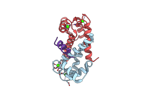
Method: X-RAY DIFFRACTION Resolution:2.25 Å Release Date: 2013-01-30 Classification: METAL BINDING PROTEIN Ligands: CA |
 |
Organism: Homo sapiens
Method: X-RAY DIFFRACTION Resolution:1.54 Å Release Date: 2012-07-11 Classification: METAL BINDING PROTEIN Ligands: CA |
 |
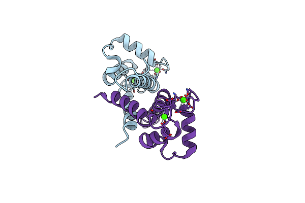
Structure Of The Tfp-Ca2+-Bound Activated Form Of The S100A4 Metastasis Factor
Organism: Homo sapiens
Method: X-RAY DIFFRACTION Resolution:2.30 Å Release Date: 2010-05-26 Classification: METAL BINDING PROTEIN Ligands: TFP, CA |
 |
Organism: Homo sapiens
Method: X-RAY DIFFRACTION Release Date: 2010-05-12 Classification: Calcium Binding Protein Ligands: P77, CA, DIO |
 |
Organism: Homo sapiens
Method: X-RAY DIFFRACTION Resolution:1.63 Å Release Date: 2008-02-26 Classification: METAL BINDING PROTEIN Ligands: CA |
 |
Organism: Homo sapiens
Method: SOLUTION NMR Release Date: 2002-10-30 Classification: METAL BINDING PROTEIN |
 |
Organism: Escherichia coli
Method: X-RAY DIFFRACTION Resolution:2.00 Å Release Date: 2001-08-22 Classification: OXIDOREDUCTASE, LYASE Ligands: FE2 |