
Search Count: 50
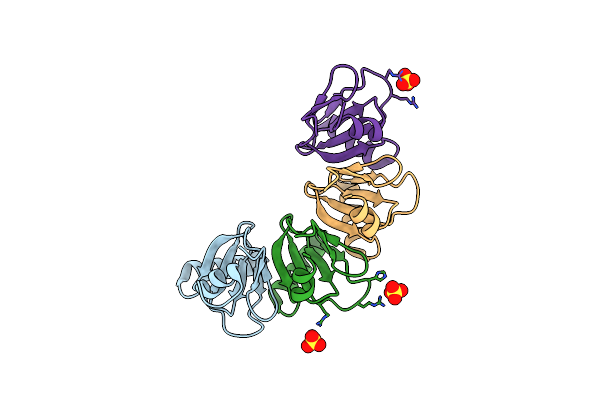 |
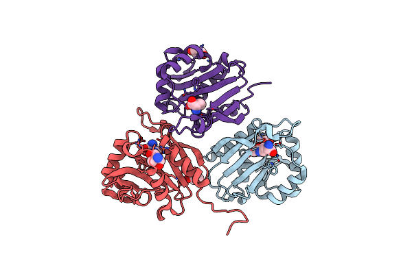
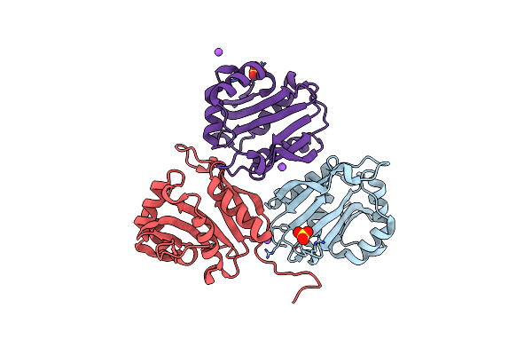
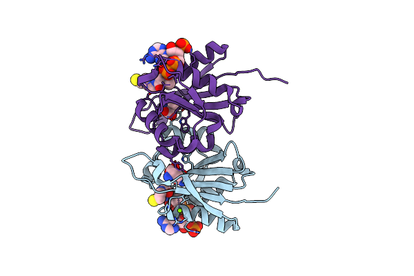
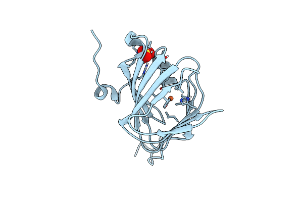
Crystal Structure Of The Dna-Binding Protein Rema From Geobacillus Thermodenitrificans
Organism: Geobacillus thermodenitrificans ng80-2
Method: X-RAY DIFFRACTION Resolution:2.29 Å Release Date: 2021-08-25 Classification: DNA BINDING PROTEIN Ligands: SO4 |
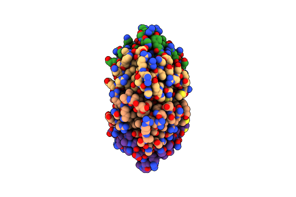 |
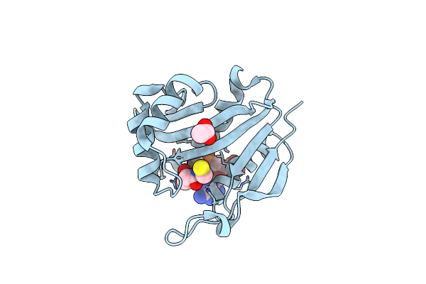
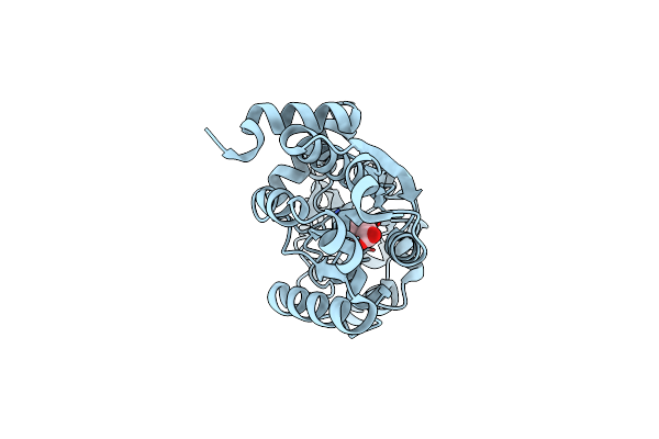
Crystal Structure Of A R18W Mutant Of The Dna-Binding Protein Rema From Geobacillus Thermodenitrificans
Organism: Geobacillus thermodenitrificans (strain ng80-2)
Method: X-RAY DIFFRACTION Resolution:2.60 Å Release Date: 2021-08-25 Classification: DNA BINDING PROTEIN |
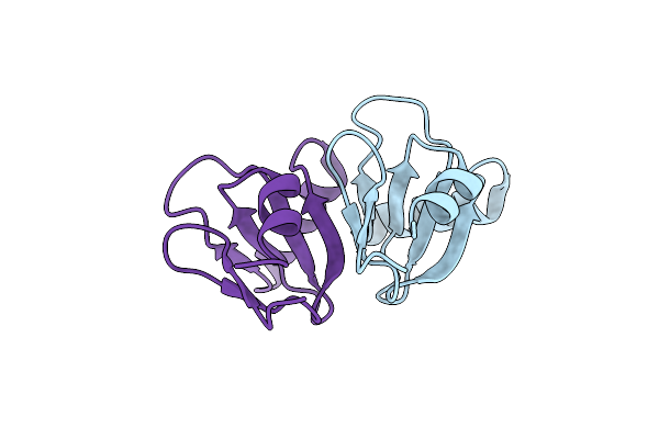 |
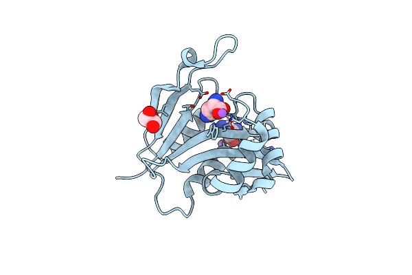
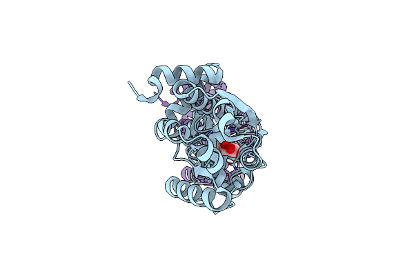
Crystal Structure Of A R51 R53 Double Mutant Of The Dna-Binding Protein Rema From Geobacillus Thermodenitrificans
Organism: Geobacillus thermodenitrificans (strain ng80-2)
Method: X-RAY DIFFRACTION Resolution:1.80 Å Release Date: 2021-08-25 Classification: DNA BINDING PROTEIN |
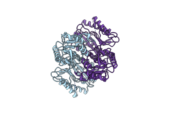 |
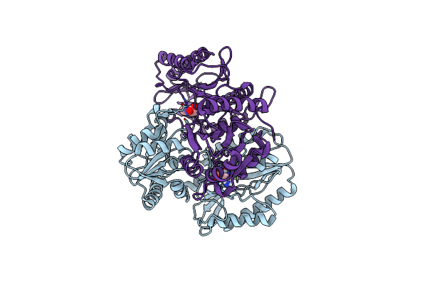
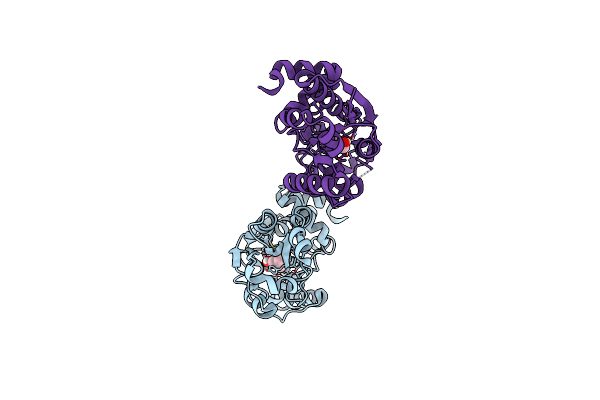
Apo Structure Of The Ectoine Utilization Protein Eutd (Doea) From Halomonas Elongata
Organism: Halomonas elongata
Method: X-RAY DIFFRACTION Resolution:2.15 Å Release Date: 2020-05-20 Classification: HYDROLASE |
 |
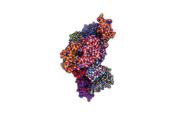
Substrate Bound Structure Of The Ectoine Utilization Protein Eutd (Doea) From Halomonas Elongata
Organism: Halomonas elongata
Method: X-RAY DIFFRACTION Resolution:2.25 Å Release Date: 2020-05-20 Classification: HYDROLASE Ligands: P4B, 4CS |
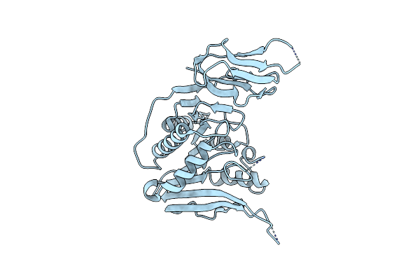 |
Apo Structure Of The Ectoine Utilization Protein Eute (Doeb) From Ruegeria Pomeroyi
Organism: Ruegeria pomeroyi (strain atcc 700808 / dsm 15171 / dss-3)
Method: X-RAY DIFFRACTION Resolution:2.00 Å Release Date: 2020-05-20 Classification: HYDROLASE |
 |
Product Bound Structure Of The Ectoine Utilization Protein Eute (Doeb) From Ruegeria Pomeroyi
Organism: Ruegeria pomeroyi (strain atcc 700808 / dsm 15171 / dss-3)
Method: X-RAY DIFFRACTION Resolution:2.50 Å Release Date: 2020-05-20 Classification: HYDROLASE Ligands: ZN, DAB, ACT |
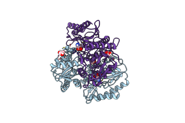 |
Product Bound Structure Of The Ectoine Utilization Protein Eutd (Doea) From Halomonas Elongata
Organism: Halomonas elongata
Method: X-RAY DIFFRACTION Resolution:2.40 Å Release Date: 2020-05-20 Classification: HYDROLASE Ligands: GOL, P4B |
 |
Diaminobutyrate Acetyltransferase Ecta From Paenibacillus Lautus In Complex With Its Product Adaba
Organism: Geobacillus sp. (strain y412mc10)
Method: X-RAY DIFFRACTION Resolution:2.20 Å Release Date: 2020-01-29 Classification: TRANSFERASE Ligands: GOL, 9YT, TRS |
 |
Diaminobutyrate Acetyltransferase Ecta From Paenibacillus Lautus In Complex With Coenzyme A
Organism: Geobacillus sp. (strain y412mc10)
Method: X-RAY DIFFRACTION Resolution:1.50 Å Release Date: 2020-01-29 Classification: TRANSFERASE Ligands: COA, ACT |
 |
Diaminobutyrate Acetyltransferase Ecta From Paenibacillus Lautus In Complex With Its Substrate L-2,4-Diaminobutyric Acid (Dab)
Organism: Geobacillus sp. (strain y412mc10)
Method: X-RAY DIFFRACTION Resolution:1.53 Å Release Date: 2020-01-29 Classification: TRANSFERASE Ligands: DAB, NA, GOL |
 |
Organism: Geobacillus sp. (strain y412mc10)
Method: X-RAY DIFFRACTION Resolution:2.20 Å Release Date: 2020-01-29 Classification: TRANSFERASE |
 |
Diaminobutyrate Acetyltransferase Ecta From Paenibacillus Lautus In Complex With Its Substrate L-2,4-Diaminobutyric Acid (Dab) And Coenzyme A
Organism: Geobacillus sp. (strain y412mc10)
Method: X-RAY DIFFRACTION Resolution:1.20 Å Release Date: 2020-01-29 Classification: TRANSFERASE Ligands: COA, DAB, MG |
 |
Crystal Structure Of The Beta-Hydroxyaspartate Aldolase Of Paracoccus Denitrificans
Organism: Paracoccus denitrificans
Method: X-RAY DIFFRACTION Resolution:1.70 Å Release Date: 2019-08-14 Classification: LYASE Ligands: PLP, MG |
 |
Crystal Structure Of The Iminosuccinate Reductase Of Paracoccus Denitrificans In Complex With Nad+
Organism: Paracoccus denitrificans (strain pd 1222)
Method: X-RAY DIFFRACTION Resolution:2.56 Å Release Date: 2019-08-14 Classification: OXIDOREDUCTASE Ligands: NAD, TB, 7MT |
 |
Organism: Bacillus subtilis
Method: X-RAY DIFFRACTION Resolution:1.42 Å Release Date: 2018-11-21 Classification: TRANSPORT PROTEIN Ligands: BET |
 |
Organism: Bacillus subtilis subsp. subtilis str. 168
Method: X-RAY DIFFRACTION Resolution:1.60 Å Release Date: 2018-11-21 Classification: TRANSPORT PROTEIN Ligands: DQY |
 |
Organism: Bacillus subtilis
Method: X-RAY DIFFRACTION Resolution:1.50 Å Release Date: 2018-11-21 Classification: TRANSPORT PROTEIN Ligands: 152 |
 |
Organism: Bacillus subtilis (strain 168)
Method: X-RAY DIFFRACTION Resolution:1.50 Å Release Date: 2018-11-21 Classification: TRANSPORT PROTEIN Ligands: CHT |
 |
Organism: Paenibacillus lautus
Method: X-RAY DIFFRACTION Resolution:1.52 Å Release Date: 2018-08-22 Classification: METAL BINDING PROTEIN Ligands: FE, SO4 |