
Search Count: 127
 |
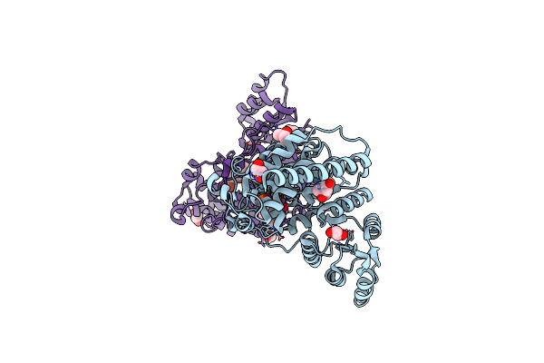
Organism: Escherichia coli
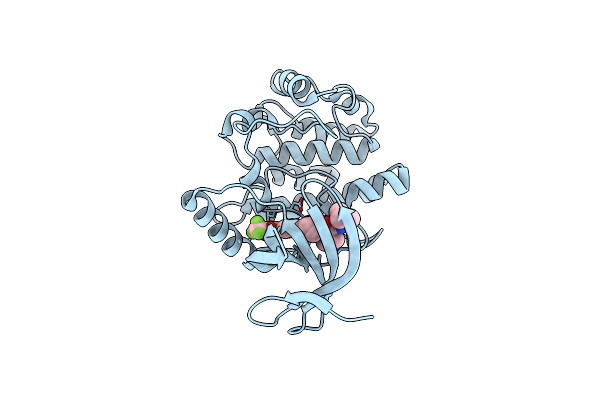
Method: X-RAY DIFFRACTION Resolution:1.60 Å Release Date: 2024-09-18 Classification: HYDROLASE Ligands: MN, A1H5P |
 |
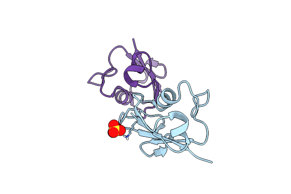
Organism: Klebsiella pneumoniae
Method: X-RAY DIFFRACTION Resolution:2.20 Å Release Date: 2024-09-18 Classification: HYDROLASE Ligands: MN, A1H55 |
 |
Organism: Escherichia coli
Method: X-RAY DIFFRACTION Resolution:1.35 Å Release Date: 2024-04-17 Classification: HYDROLASE Ligands: MN, LP5 |
 |
Organism: Klebsiella pneumoniae
Method: X-RAY DIFFRACTION Resolution:1.95 Å Release Date: 2024-04-17 Classification: HYDROLASE Ligands: MN, VTF |
 |
Organism: Klebsiella pneumoniae
Method: X-RAY DIFFRACTION Resolution:1.95 Å Release Date: 2024-04-17 Classification: HYDROLASE Ligands: MN, VN6 |
 |
Organism: Escherichia coli
Method: X-RAY DIFFRACTION Resolution:1.90 Å Release Date: 2024-04-17 Classification: HYDROLASE Ligands: MN, VQ9 |
 |
Organism: Klebsiella pneumoniae
Method: X-RAY DIFFRACTION Resolution:2.00 Å Release Date: 2024-04-17 Classification: HYDROLASE Ligands: MN, VTK |
 |
Organism: Drosophila melanogaster
Method: X-RAY DIFFRACTION Resolution:1.52 Å Release Date: 2021-08-04 Classification: SIGNALING PROTEIN Ligands: CA, FUC, BGC, NAG |
 |
Organism: Drosophila melanogaster
Method: X-RAY DIFFRACTION Resolution:3.00 Å Release Date: 2021-08-04 Classification: SIGNALING PROTEIN Ligands: NAG |
 |
Organism: Drosophila melanogaster
Method: X-RAY DIFFRACTION Resolution:2.03 Å Release Date: 2021-08-04 Classification: SIGNALING PROTEIN Ligands: CA, NAG |
 |
Organism: Sulfolobus acidocaldarius
Method: X-RAY DIFFRACTION Resolution:3.70 Å Release Date: 2020-08-05 Classification: HYDROLASE |
 |
Organism: Rattus norvegicus
Method: X-RAY DIFFRACTION Resolution:1.65 Å Release Date: 2016-09-07 Classification: TRANSFERASE/TRANSFERASE INHIBITOR Ligands: 5FJ |
 |
Structure Of Plasmodium Falciparum Dxr In Complex With A Beta-Substituted Fosmidomycin Analogue, Lc51 And Manganese
Organism: Plasmodium falciparum (isolate 3d7)
Method: X-RAY DIFFRACTION Resolution:1.40 Å Release Date: 2016-08-24 Classification: OXIDOREDUCTASE Ligands: LC5, MN, GOL, FMT |
 |
Structure Of Plasmodium Falciparum Dxr In Complex With A Beta-Substituted Fosmidomycin Analogue, Lc52 And Manganese
Organism: Plasmodium falciparum (isolate 3d7)
Method: X-RAY DIFFRACTION Resolution:1.70 Å Release Date: 2016-08-24 Classification: OXIDOREDUCTASE Ligands: 6J8, MN, GOL, EDO |
 |
Structure Of Plasmodium Falciparum Dxr In Complex With A Beta-Substituted Fosmidomycin Analogue, Lc55 And Manganese
Organism: Plasmodium falciparum (isolate 3d7)
Method: X-RAY DIFFRACTION Resolution:1.65 Å Release Date: 2016-08-24 Classification: OXIDOREDUCTASE Ligands: 6JB, MN, GOL, EDO |
 |
Structure Of Plasmodium Falciparum Dxr In Complex With A Beta-Substituted Fosmidomycin Analogue, Lc57 And Manganese
Organism: Plasmodium falciparum 3d7
Method: X-RAY DIFFRACTION Resolution:1.70 Å Release Date: 2016-08-24 Classification: OXIDOREDUCTASE Ligands: LC7, MN, GOL, 6LB, EDO |
 |
Structure Of Plasmodium Falciparum Dxr In Complex With A Beta-Substituted Fosmidomycin Analogue, Lc50 And Manganese
Organism: Plasmodium falciparum (isolate 3d7)
Method: X-RAY DIFFRACTION Resolution:1.55 Å Release Date: 2016-08-24 Classification: OXIDOREDUCTASE Ligands: L50, MN, MPD, EDO |
 |
Structure Of Plasmodium Falciparum Dxr In Complex With A Beta-Substituted Fosmidomycin Analogue, Lc54 And Manganese
Organism: Plasmodium falciparum (isolate 3d7)
Method: X-RAY DIFFRACTION Resolution:1.60 Å Release Date: 2016-08-24 Classification: OXIDOREDUCTASE Ligands: L54, MN, GOL, EDO |
 |
Structure Of Plasmodium Falciparum Dxr In Complex With A Beta-Substituted Fosmidomycin Analogue, Lc56 And Manganese
Organism: Plasmodium falciparum 3d7
Method: X-RAY DIFFRACTION Resolution:1.80 Å Release Date: 2016-08-24 Classification: OXIDOREDUCTASE Ligands: L56, MN, GOL, EDO, 6LR |
 |
Organism: Sulfolobus solfataricus
Method: X-RAY DIFFRACTION Resolution:2.20 Å Release Date: 2015-09-23 Classification: SIGNALING PROTEIN Ligands: SO4 |