
Search Count: 165
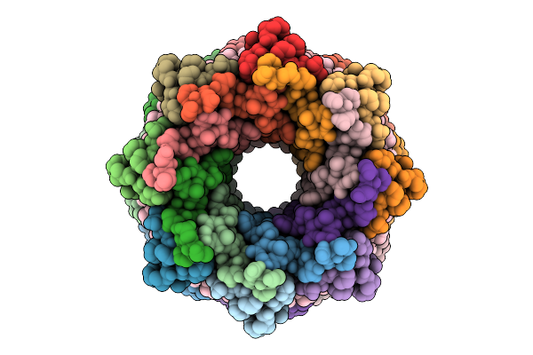 |
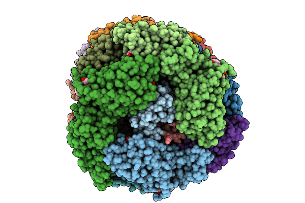
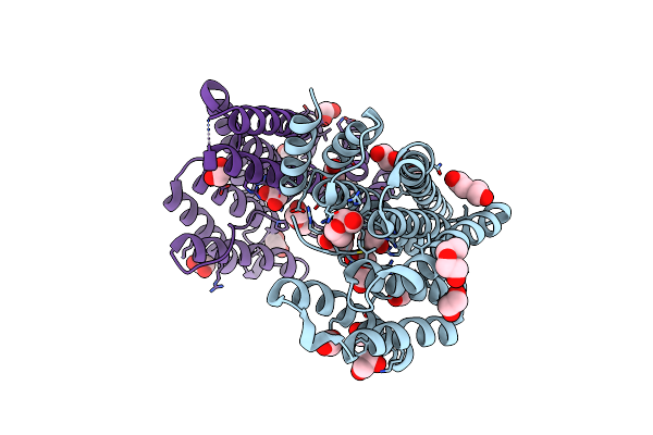
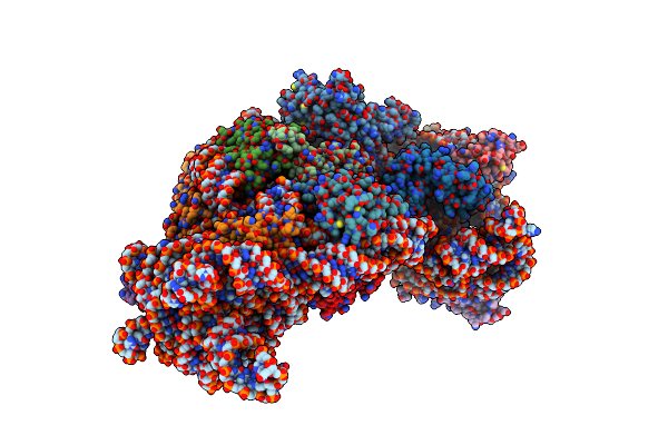
Lh2 Complex From Ectothiorhodospira Haloalkaliphila With Inhibited Carotenoid Biosynthesis
Organism: Ectothiorhodospira haloalkaliphila atcc 51935
Method: ELECTRON MICROSCOPY Release Date: 2025-10-22 Classification: PHOTOSYNTHESIS Ligands: BCL, A1MBA |
 |
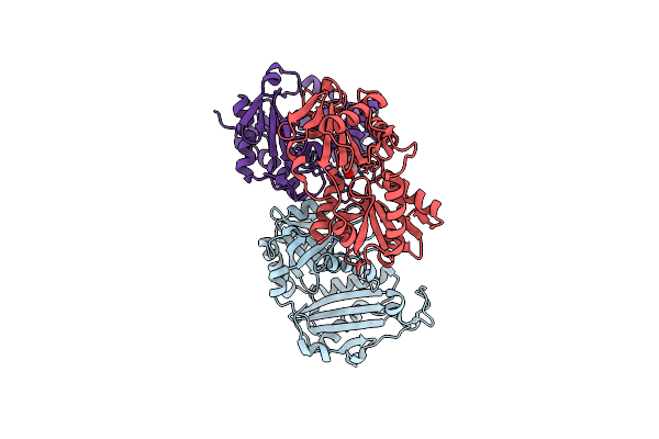
Organism: Drosophila melanogaster
Method: X-RAY DIFFRACTION Release Date: 2025-10-08 Classification: TRANSCRIPTION |
 |
Organism: Gloeobacter violaceus pcc 7421
Method: ELECTRON MICROSCOPY Release Date: 2025-09-24 Classification: PHOTOSYNTHESIS Ligands: CYC |
 |
Organism: Gloeobacter violaceus pcc 7421
Method: ELECTRON MICROSCOPY Release Date: 2025-09-24 Classification: PHOTOSYNTHESIS Ligands: CYC |
 |
Organism: Gloeobacter violaceus pcc 7421
Method: ELECTRON MICROSCOPY Release Date: 2025-09-24 Classification: PHOTOSYNTHESIS Ligands: CYC |
 |
Organism: Gloeobacter violaceus pcc 7421
Method: ELECTRON MICROSCOPY Release Date: 2025-09-24 Classification: PHOTOSYNTHESIS Ligands: CYC |
 |
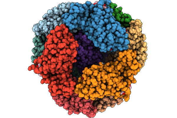
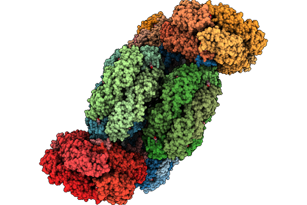
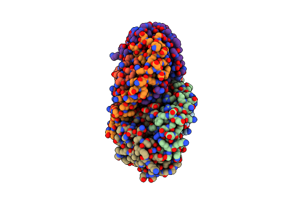
Phycobilisome Allophycocyanin Hexamer C From Gloeobacter Violaceus Pcc 7421
Organism: Gloeobacter violaceus pcc 7421
Method: ELECTRON MICROSCOPY Release Date: 2025-09-24 Classification: PHOTOSYNTHESIS Ligands: CYC |
 |
Organism: Gloeobacter violaceus pcc 7421
Method: ELECTRON MICROSCOPY Release Date: 2025-09-24 Classification: PHOTOSYNTHESIS Ligands: CYC |
 |
Crystal Structure Of The Genetically-Encoded Green Calcium Sensor Icbtnc2 In Its Calcium Bound-State
Organism: Synthetic construct
Method: X-RAY DIFFRACTION Release Date: 2025-09-17 Classification: FLUORESCENT PROTEIN Ligands: PEG, CA, CL |
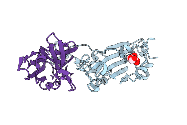 |
Crystal Structure Of The Wuhan Sars-Cov-2 Spike Rbd (319-541) Complexed With 1P1B10 Nanobody
Organism: Severe acute respiratory syndrome coronavirus 2, Camelus bactrianus
Method: X-RAY DIFFRACTION Release Date: 2025-07-16 Classification: VIRAL PROTEIN/IMMUNE SYSTEM Ligands: NAG, NA |
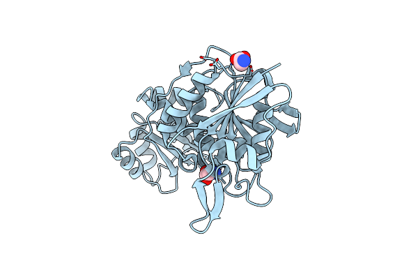 |
Organism: Thermococcus sibiricus
Method: X-RAY DIFFRACTION Release Date: 2025-06-11 Classification: HYDROLASE Ligands: GLY |
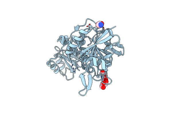 |
Crystal Structure Of L-Asparaginase From Thermococcus Sibiricus Double Mutation D54G/T56Q
Organism: Thermococcus sibiricus
Method: X-RAY DIFFRACTION Release Date: 2025-06-11 Classification: HYDROLASE Ligands: PEG, GOL, GLY |
 |
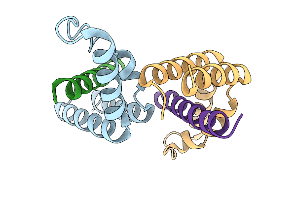
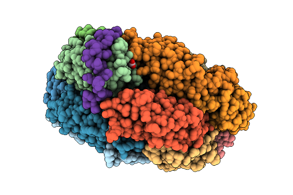
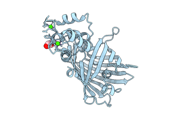
14-3-3 Zeta Chimera With The S202R Peptide Of Sars-Cov-2 N (Residues 200-213)
Organism: Homo sapiens, Severe acute respiratory syndrome coronavirus 2
Method: X-RAY DIFFRACTION Resolution:1.80 Å Release Date: 2025-05-21 Classification: SIGNALING PROTEIN Ligands: EDO, PEG |
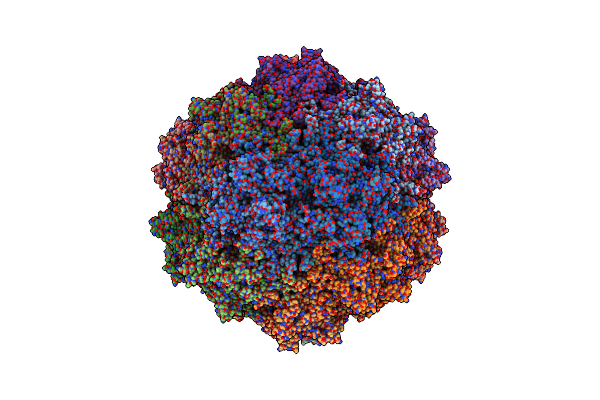 |
Organism: Adeno-associated virus 5
Method: ELECTRON MICROSCOPY Resolution:2.55 Å Release Date: 2025-04-30 Classification: VIRUS |
 |
Crystal Structure Of The Soluble Green Pigment Protein From Tettigonia Cantans
Organism: Tettigonia cantans
Method: X-RAY DIFFRACTION Release Date: 2025-04-23 Classification: TRANSPORT PROTEIN Ligands: LUT, AZI, PLC, A1L6M |
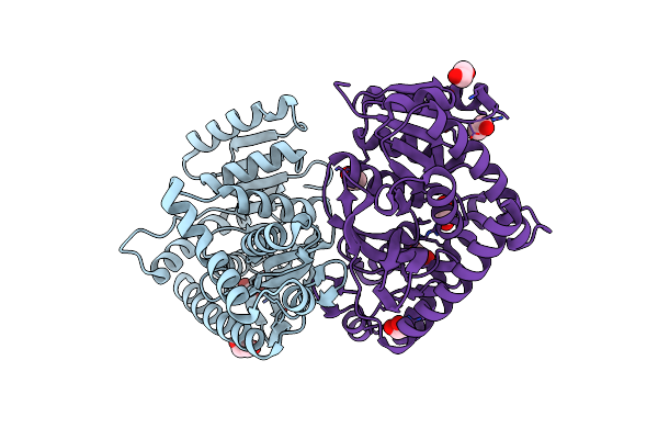 |
Crystal Structure L-Lactate Dehydrogenase From Lactobacillus Reuteri In Its Apoform
Organism: Limosilactobacillus reuteri
Method: X-RAY DIFFRACTION Resolution:2.15 Å Release Date: 2025-04-02 Classification: OXIDOREDUCTASE Ligands: EDO |
 |
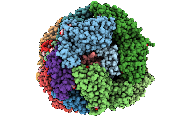
Organism: Escherichia coli
Method: ELECTRON MICROSCOPY Release Date: 2025-02-05 Classification: RIBOSOME |
 |
Organism: Escherichia coli
Method: ELECTRON MICROSCOPY Release Date: 2025-02-05 Classification: RIBOSOME |
 |
Crystal Structure Of Aminotransferase-Like Protein From Variovorax Paradoxus Mutant N174K
Organism: Variovorax paradoxus b4
Method: X-RAY DIFFRACTION Resolution:2.20 Å Release Date: 2025-01-29 Classification: TRANSFERASE Ligands: PEG |
 |
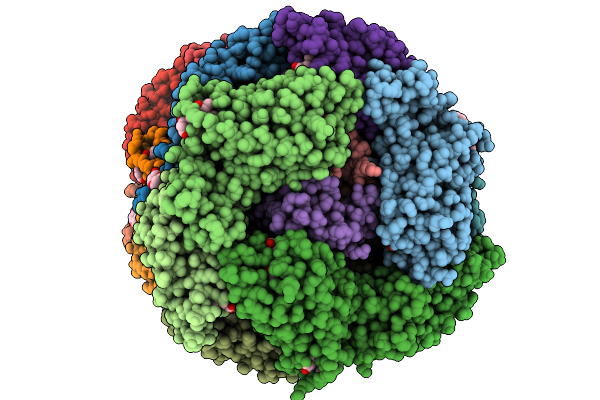
Lh2 Complex From Ectothiorhodospira Haloalkaliphila At Near-Atomic Resolution
Organism: Ectothiorhodospira haloalkaliphila atcc 51935
Method: ELECTRON MICROSCOPY Resolution:1.70 Å Release Date: 2025-01-01 Classification: PHOTOSYNTHESIS Ligands: BCL, A1L0S |