
Search Count: 21
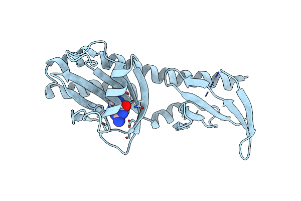 |
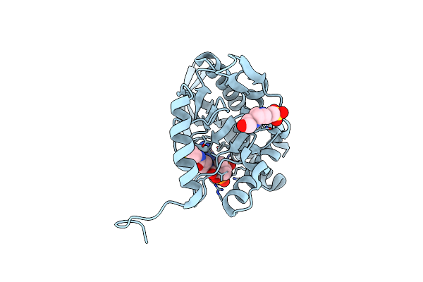
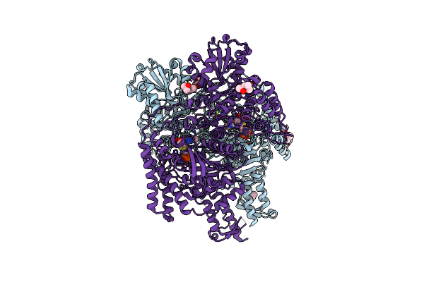
Crystal Structure Of The Ligand Binding Domain Of The Halomonas Titanicae Chemoreceptor Htc10 In Complex With Guanine
Organism: Halomonas titanicae
Method: X-RAY DIFFRACTION Resolution:3.60 Å Release Date: 2024-11-20 Classification: SIGNALING PROTEIN Ligands: GUN |
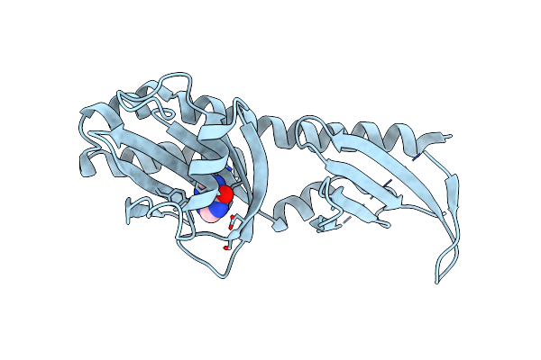 |
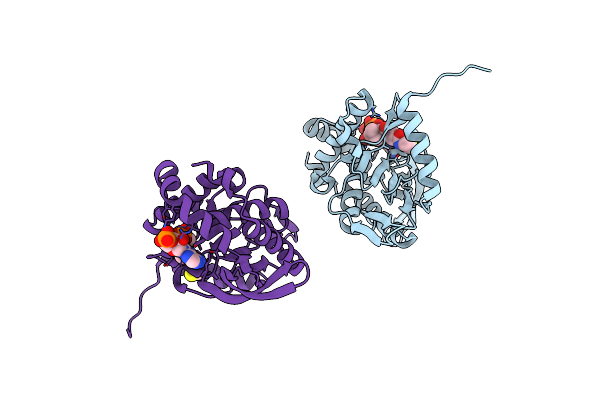
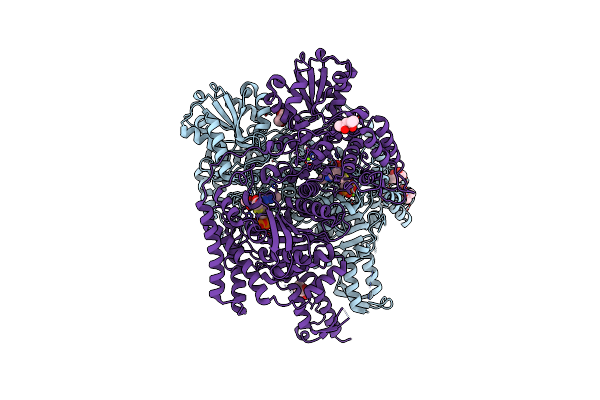
Crystal Structure Of The Ligand Binding Domain Of The Halomonas Titanicae Chemoreceptor Htc10 In Complex With Hypoxanthine
Organism: Halomonas titanicae
Method: X-RAY DIFFRACTION Resolution:3.63 Å Release Date: 2024-11-20 Classification: SIGNALING PROTEIN Ligands: HPA |
 |
High-Resolution Crystal Structure Of The Ligand Binding Domain Of The Halomonas Titanicae Chemoreceptor Htc10 In Complex With Guanine
Organism: Halomonas titanicae
Method: X-RAY DIFFRACTION Resolution:2.10 Å Release Date: 2024-11-20 Classification: SIGNALING PROTEIN Ligands: GUN |
 |
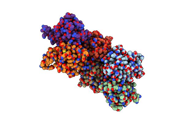
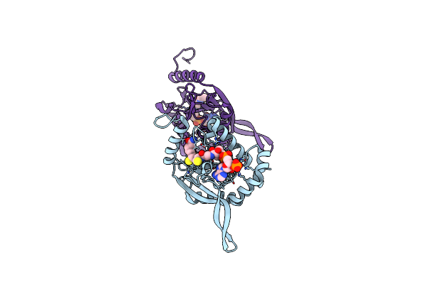
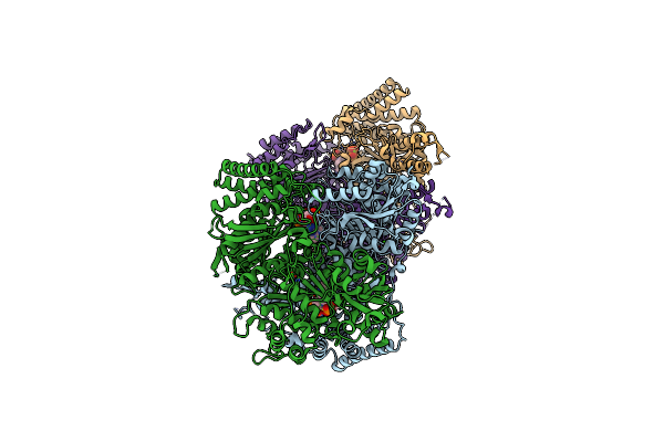
Crystal Structure Of Full-Length, Homohexameric 2-Oxoglutarate Dehydrogenase Kgd From Mycobacterium Smegmatis In Complex With Gara
Organism: Mycolicibacterium smegmatis mc2 155
Method: X-RAY DIFFRACTION Resolution:4.56 Å Release Date: 2023-08-16 Classification: OXIDOREDUCTASE Ligands: MG, CA, TPP |
 |
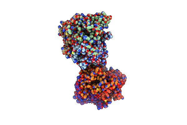
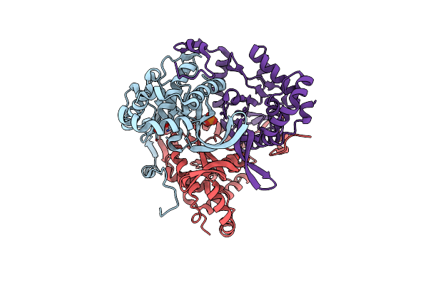
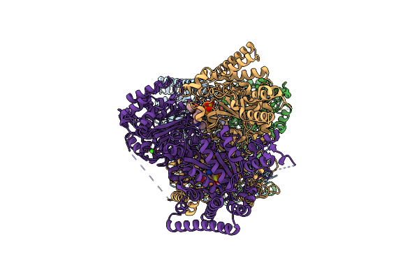
Crystal Structure Of The Homohexameric 2-Oxoglutarate Dehydrogenase Odha From Corynebacterium Glutamicum
Organism: Corynebacterium glutamicum atcc 13032
Method: X-RAY DIFFRACTION Resolution:2.46 Å Release Date: 2023-08-16 Classification: OXIDOREDUCTASE Ligands: EPE, COA, ACO, TPP, MG |
 |
Single Particle Cryo-Em Structure Of The Homohexameric 2-Oxoglutarate Dehydrogenase Odha From Corynebacterium Glutamicum
Organism: Corynebacterium glutamicum atcc 13032
Method: ELECTRON MICROSCOPY Resolution:2.17 Å Release Date: 2023-08-16 Classification: OXIDOREDUCTASE Ligands: MG, ACO, TPP |
 |
Single Particle Cryo-Em Structure Of Homohexameric 2-Oxoglutarate Dehydrogenase Odha From Corynebacterium Glutamicum With Coenzyme A Bound To The E2O Domain
Organism: Corynebacterium glutamicum atcc 13032
Method: ELECTRON MICROSCOPY Resolution:2.17 Å Release Date: 2023-08-16 Classification: OXIDOREDUCTASE Ligands: MG, COA, ACO, TPP |
 |
Single Particle Cryo-Em Structure Of Homohexameric 2-Oxoglutarate Dehydrogenase Odha From Corynebacterium Glutamicum In Complex With The Product Succinyl-Coa
Organism: Corynebacterium glutamicum atcc 13032
Method: ELECTRON MICROSCOPY Release Date: 2023-08-16 Classification: OXIDOREDUCTASE Ligands: MG, ACO, TPP, SCA |
 |
Single Particle Cryo-Em Structure Of Homohexameric 2-Oxoglutarate Dehydrogenase Odha From Corynebacterium Glutamicum Following Reaction With The 2-Oxoglutarate Analogue Succinyl Phosphonate
Organism: Corynebacterium glutamicum atcc 13032
Method: ELECTRON MICROSCOPY Resolution:2.26 Å Release Date: 2023-08-16 Classification: OXIDOREDUCTASE Ligands: MG, ACO, QSP |
 |
Single Particle Cryo-Em Structure Of The Complex Between Corynebacterium Glutamicum Homohexameric 2-Oxoglutarate Dehydrogenase Odha And The Fha-Protein Inhibitor Odhi
Organism: Corynebacterium glutamicum atcc 13032
Method: ELECTRON MICROSCOPY Resolution:2.29 Å Release Date: 2023-08-16 Classification: OXIDOREDUCTASE Ligands: TPP, MG |
 |
Crystal Structure Of The Catalyic Domain Of Corynebacterium Glutamicum Acetyltransferase Acef (E2P).
Organism: Corynebacterium glutamicum (strain atcc 13032 / dsm 20300 / jcm 1318 / lmg 3730 / ncimb 10025)
Method: X-RAY DIFFRACTION Resolution:1.93 Å Release Date: 2021-08-18 Classification: TRANSFERASE |
 |
Crystal Structure Of The Catalytic Domain Of Corynebacterium Glutamicum Acetyltransferase Acef (E2P) In Complex With Oxidized Coa.
Organism: Corynebacterium glutamicum (strain atcc 13032 / dsm 20300 / jcm 1318 / lmg 3730 / ncimb 10025)
Method: X-RAY DIFFRACTION Resolution:1.35 Å Release Date: 2021-08-18 Classification: TRANSFERASE Ligands: CAO, EPE |
 |
Crystal Structure Of The Catalytic Domain Of C. Glutamicum Acef (E2P) In Ternary Complex With Coa And Dihydrolipoamide.
Organism: Corynebacterium glutamicum atcc 13032
Method: X-RAY DIFFRACTION Resolution:2.09 Å Release Date: 2021-08-18 Classification: TRANSFERASE Ligands: COA, LPM |
 |
Crystal Structure Of The Catalytic Domain Plus N-Terminal Linker Of The Acetyltransferase Acef (E2P) From Corynebacterium Glutamicum.
Organism: Corynebacterium glutamicum (strain atcc 13032 / dsm 20300 / jcm 1318 / lmg 3730 / ncimb 10025)
Method: X-RAY DIFFRACTION Resolution:2.23 Å Release Date: 2021-08-18 Classification: TRANSFERASE Ligands: GOL, PO4 |
 |
Crystal Structure Of The Catalytic Domain Of Corynebacterium Mustelae Predicted Acetyltransferase Acef (E2P).
Organism: Corynebacterium mustelae
Method: X-RAY DIFFRACTION Resolution:2.50 Å Release Date: 2021-08-18 Classification: TRANSFERASE Ligands: COA |
 |
Crystal Structure Of The Cubic Catalytic Core Of The Mycobacterium Tuberculosis Branched-Chain Alphaketoacid Acyltransferase Component (E2B).
Organism: Mycobacterium tuberculosis h37rv
Method: X-RAY DIFFRACTION Resolution:1.50 Å Release Date: 2021-08-18 Classification: TRANSFERASE Ligands: ACT, IMD |
 |
Crystal Structure Of The Suca Domain Of Mycobacterium Smegmatis Kgd Cocrystallized With Succinylphosphonate
Organism: Mycobacterium smegmatis (strain atcc 700084 / mc(2)155)
Method: X-RAY DIFFRACTION Resolution:1.67 Å Release Date: 2019-09-11 Classification: OXIDOREDUCTASE Ligands: QSP, MG, MPD |
 |
Crystal Structure Of The Suca Domain Of Mycobacterium Smegmatis Kgd Cocrystallized With Succinylphosphonate Phosphonoethyl Ester (Pesp)
Organism: Mycobacterium smegmatis (strain atcc 700084 / mc(2)155)
Method: X-RAY DIFFRACTION Resolution:1.70 Å Release Date: 2019-09-11 Classification: OXIDOREDUCTASE Ligands: ZP1, MG, MPD |
 |
Crystal Structure Of The Suca Domain Of Mycobacterium Smegmatis Kgd After Soaking With Succinylphosphonate
Organism: Mycobacterium smegmatis (strain atcc 700084 / mc(2)155)
Method: X-RAY DIFFRACTION Resolution:1.96 Å Release Date: 2019-09-11 Classification: OXIDOREDUCTASE Ligands: QSP, MG, CA |
 |
Crystal Structure Of The Suca Domain Of Mycobacterium Smegmatis Kgd After Soaking With Succinylphosphonate Phosphonoethyl Ester (Pesp)
Organism: Mycobacterium smegmatis (strain atcc 700084 / mc(2)155)
Method: X-RAY DIFFRACTION Resolution:2.09 Å Release Date: 2019-09-11 Classification: OXIDOREDUCTASE Ligands: TPP, MG, CA, JQ5 |