
Search Count: 27
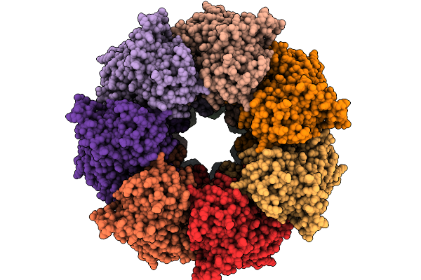 |
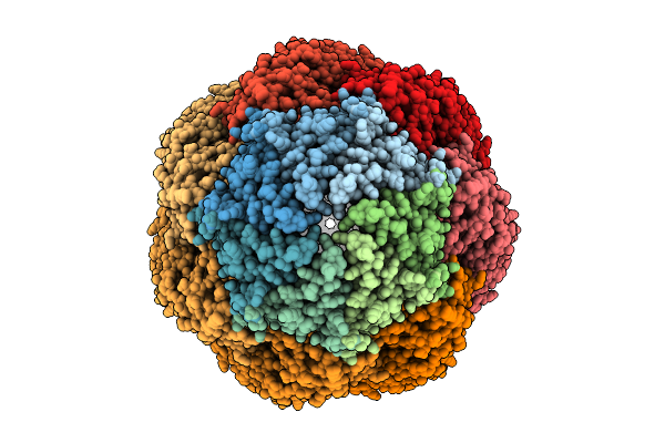
Organism: Escherichia coli
Method: ELECTRON MICROSCOPY Release Date: 2026-01-21 Classification: CHAPERONE Ligands: MG, ATP, K, A1AS6 |
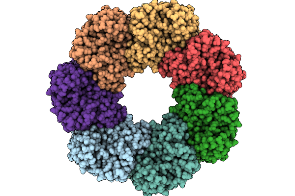 |
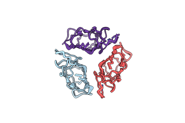
Organism: Escherichia coli
Method: ELECTRON MICROSCOPY Release Date: 2026-01-21 Classification: CHAPERONE |
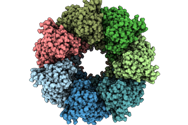 |
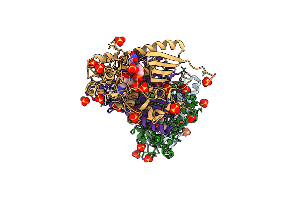
E. Coli Sr1 Single-Ring Groel Templated Into Pseudo-Double-Ring Complex With Pbz1587 Inhibitor
Organism: Escherichia coli
Method: ELECTRON MICROSCOPY Release Date: 2026-01-21 Classification: CHAPERONE Ligands: A1AS6 |
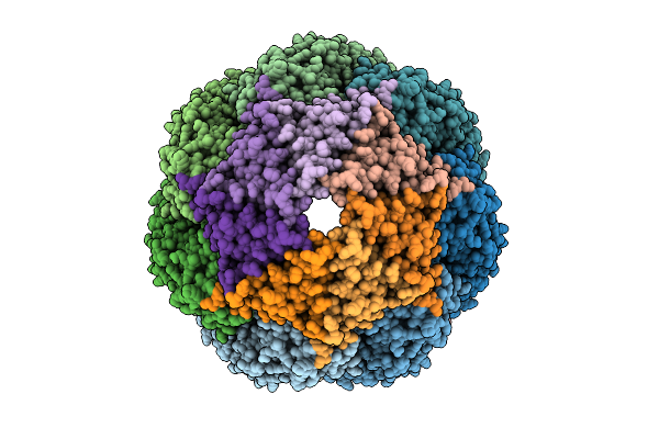 |
Organism: Escherichia coli
Method: ELECTRON MICROSCOPY Release Date: 2026-01-21 Classification: CHAPERONE Ligands: K, ATP, MG, ADP |
 |
Organism: Escherichia coli
Method: ELECTRON MICROSCOPY Release Date: 2026-01-21 Classification: CHAPERONE Ligands: ATP, MG, K |
 |
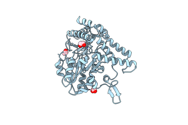
Crystal Structure Of O-Acetyl-L-Serine Sulfhydrylase A (Cysk) From Staphylococcus Aureus Nctc 8325 Complexed With A Cymr Pentapeptide
Organism: Staphylococcus aureus subsp. aureus nctc 8325, Synthetic construct
Method: X-RAY DIFFRACTION Resolution:1.80 Å Release Date: 2024-12-18 Classification: PROTEIN BINDING |
 |
Crystal Structure Of The O-Acetyl-L-Serine Sulfhydrylase A (Cysk) Holoenzyme From Staphylococcus Aureus Nctc 8325
Organism: Staphylococcus aureus subsp. aureus nctc 8325
Method: X-RAY DIFFRACTION Resolution:1.90 Å Release Date: 2024-11-13 Classification: TRANSFERASE |
 |
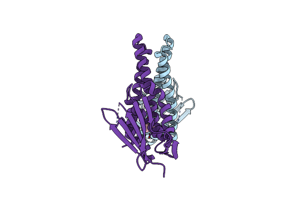
Crystal Structure Of O-Acetyl-L-Serine Sulfhydrylase A (Cysk) From Staphylococcus Aureus Nctc 8325 Complexed With A Serine Acetyltransferase (Cyse) Derived Peptide
Organism: Staphylococcus aureus subsp. aureus nctc 8325
Method: X-RAY DIFFRACTION Resolution:1.50 Å Release Date: 2024-11-13 Classification: TRANSFERASE |
 |
Crystal Structure Of O-Acetyl-L-Serine Sulfhydrylase A (Cysk) From Staphylococcus Aureus Nctc 8325 Complexed With A Transcriptional Repressor (Cymr) Derived 10 Amino Acid Peptide
Organism: Staphylococcus aureus subsp. aureus nctc 8325
Method: X-RAY DIFFRACTION Resolution:1.32 Å Release Date: 2024-11-13 Classification: TRANSFERASE |
 |
Crystal Structure Of O-Acetyl-L-Serine Sulfhydrylase A (Cysk) From Staphylococcus Aureus Nctc 8325 Complexed With A Modified Peptide Inhibitor
Organism: Staphylococcus aureus subsp. aureus nctc 8325, Synthetic construct
Method: X-RAY DIFFRACTION Resolution:2.15 Å Release Date: 2024-11-13 Classification: TRANSFERASE |
 |
Organism: Staphylococcus aureus
Method: X-RAY DIFFRACTION Resolution:1.39 Å Release Date: 2022-12-07 Classification: BIOSYNTHETIC PROTEIN Ligands: EDO |
 |
Organism: Staphylococcus aureus
Method: X-RAY DIFFRACTION Resolution:2.00 Å Release Date: 2020-04-22 Classification: MEMBRANE PROTEIN Ligands: MPD |
 |
S. Aureus Ftsz In Complex With 3-(1-(5-Bromo-4-(4-(Trifluoromethyl)Phenyl)Oxazol-2-Yl)Ethoxy)-2,6-Difluorobenzamide (Compound 2)
Organism: Staphylococcus aureus
Method: X-RAY DIFFRACTION Resolution:1.40 Å Release Date: 2020-01-15 Classification: HYDROLASE Ligands: GDP, CA, ZI1 |
 |
Organism: Staphylococcus aureus
Method: X-RAY DIFFRACTION Resolution:1.60 Å Release Date: 2020-01-15 Classification: HYDROLASE Ligands: GDP, CA, DVX |
 |
Fad Binding By Apbe Protein From Salmonella Enterica: A New Class Of Fad Binding Proteins
Organism: Salmonella enterica subsp. enterica serovar typhimurium
Method: X-RAY DIFFRACTION Resolution:2.75 Å Release Date: 2011-01-05 Classification: PROTEIN BINDING Ligands: SO4, FAD |
 |
Solution Nmr Structure Of Ydhk C-Terminal Domain From B.Subtilis, Northeast Structural Genomics Consortium Target Target Sr518
Organism: Bacillus subtilis
Method: SOLUTION NMR Release Date: 2010-07-21 Classification: STRUCTURAL GENOMICS, UNKNOWN FUNCTION |
 |
Organism: Homo sapiens
Method: SOLUTION NMR Release Date: 2008-11-25 Classification: STRUCTURAL PROTEIN |
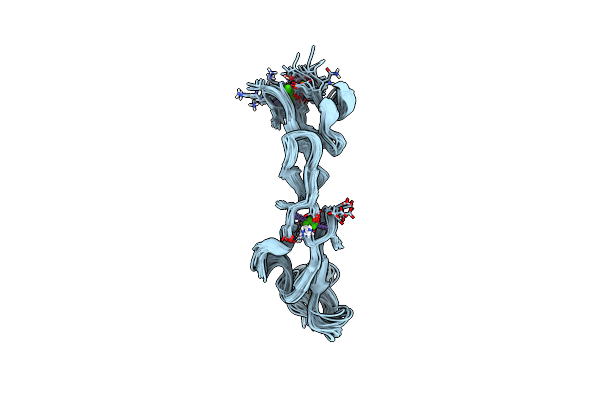 |
Nmr Study Of A Pair Of Ldl Receptor Ca2+ Binding Epidermal Growth Factor-Like Domains, 20 Structures
Organism: Homo sapiens
Method: SOLUTION NMR Release Date: 2001-07-11 Classification: CELL-SURFACE RECEPTOR Ligands: CA |
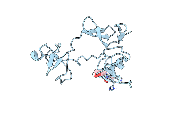 |
Solution Structure Of 6F11F22F2, A Compact Three-Module Fragment Of The Gelatin-Binding Domain Of Human Fibronectin
Organism: Homo sapiens
Method: SOLUTION NMR Release Date: 2000-10-15 Classification: CELL ADHESION Ligands: NAG |
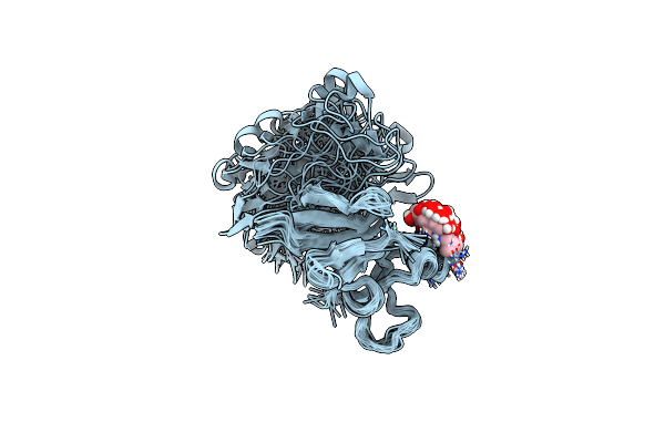 |
Solution Structure Of 6F11F22F2, A Compact Three-Module Fragment Of The Gelatin-Binding Domain Of Human Fibronectin
Organism: Homo sapiens
Method: SOLUTION NMR Release Date: 2000-10-09 Classification: CELL ADHESION Ligands: NAG |