Search Count: 17
 |
Crystal Structure Of Penicillin-Binding Protein 1 (Pbp1) From Staphylococcus Aureus
Organism: Staphylococcus aureus subsp. aureus col
Method: X-RAY DIFFRACTION Resolution:3.03 Å Release Date: 2021-11-03 Classification: HYDROLASE Ligands: EPE, CD, CL |
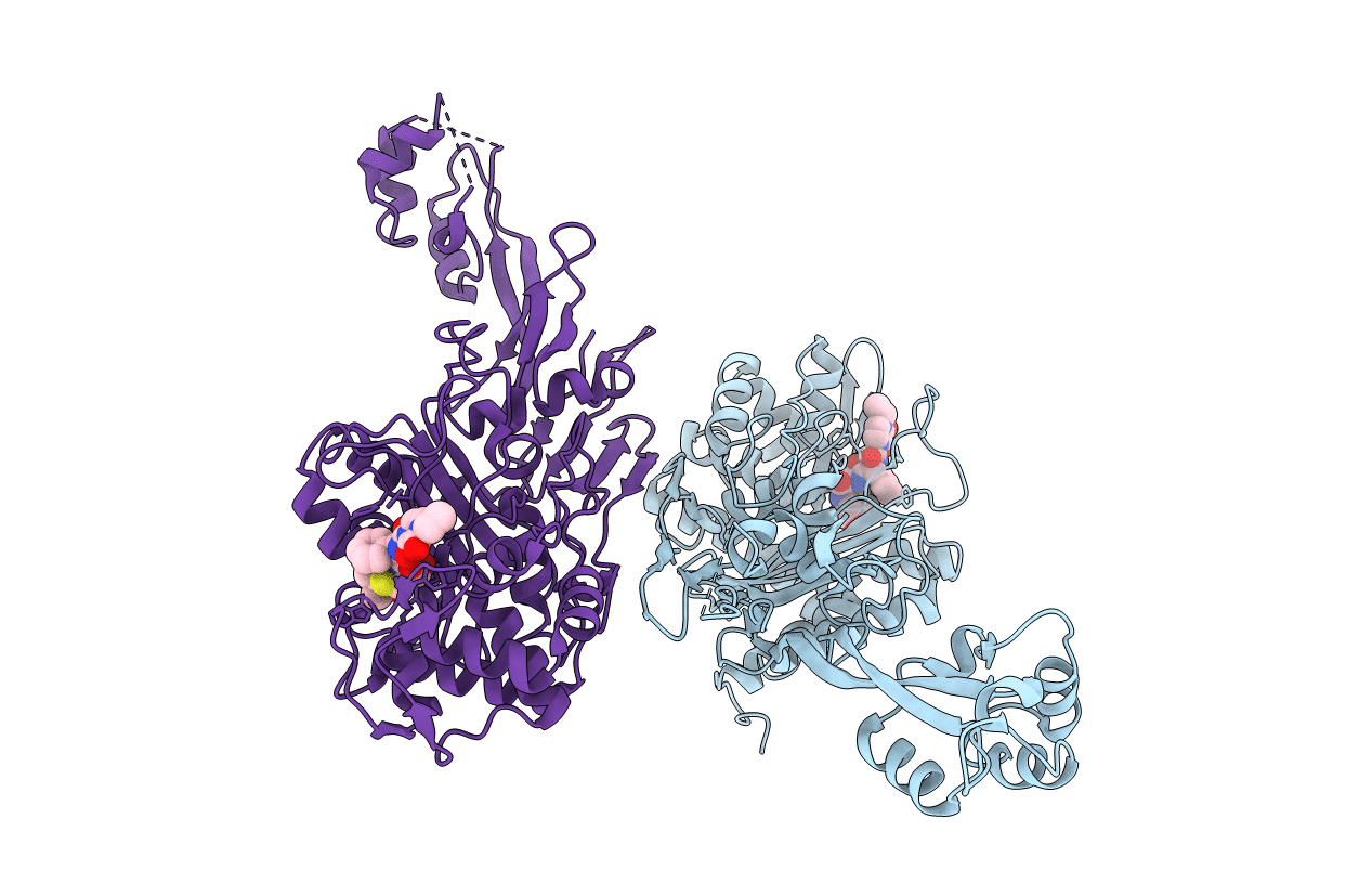 |
Crystal Structure Of Penicillin-Binding Protein 1 (Pbp1) From Staphylococcus Aureus In Complex With Piperacillin
Organism: Staphylococcus aureus subsp. aureus col
Method: X-RAY DIFFRACTION Resolution:3.03 Å Release Date: 2021-11-03 Classification: HYDROLASE Ligands: YPP |
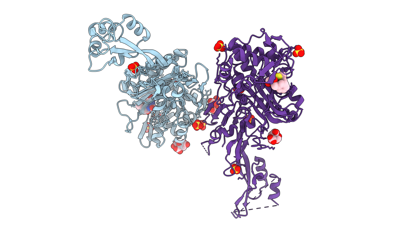 |
Crystal Structure Of Penicillin-Binding Protein 1 (Pbp1) From Staphylococcus Aureus In Complex With Penicillin G
Organism: Staphylococcus aureus subsp. aureus col
Method: X-RAY DIFFRACTION Resolution:2.59 Å Release Date: 2021-11-03 Classification: HYDROLASE Ligands: CIT, SO4, PNM |
 |
Crystal Structure Of Pasta Domains Of The Penicillin-Binding Protein 1 (Pbp1) From Staphylococcus Aureus
Organism: Staphylococcus aureus subsp. aureus col
Method: X-RAY DIFFRACTION Resolution:1.51 Å Release Date: 2021-11-03 Classification: HYDROLASE Ligands: CL |
 |
Crystal Structure Of Penicillin-Binding Protein 1 (Pbp1) From Staphylococcus Aureus In Complex With Pentaglycine
Organism: Staphylococcus aureus (strain col), Synthetic construct
Method: X-RAY DIFFRACTION Resolution:3.36 Å Release Date: 2021-11-03 Classification: HYDROLASE Ligands: CD, CL |
 |
Organism: Homo sapiens, Bos taurus
Method: X-RAY DIFFRACTION Resolution:2.44 Å Release Date: 2020-04-15 Classification: TRANSFERASE/SIGNALING PROTEIN Ligands: Q1Y, MG |
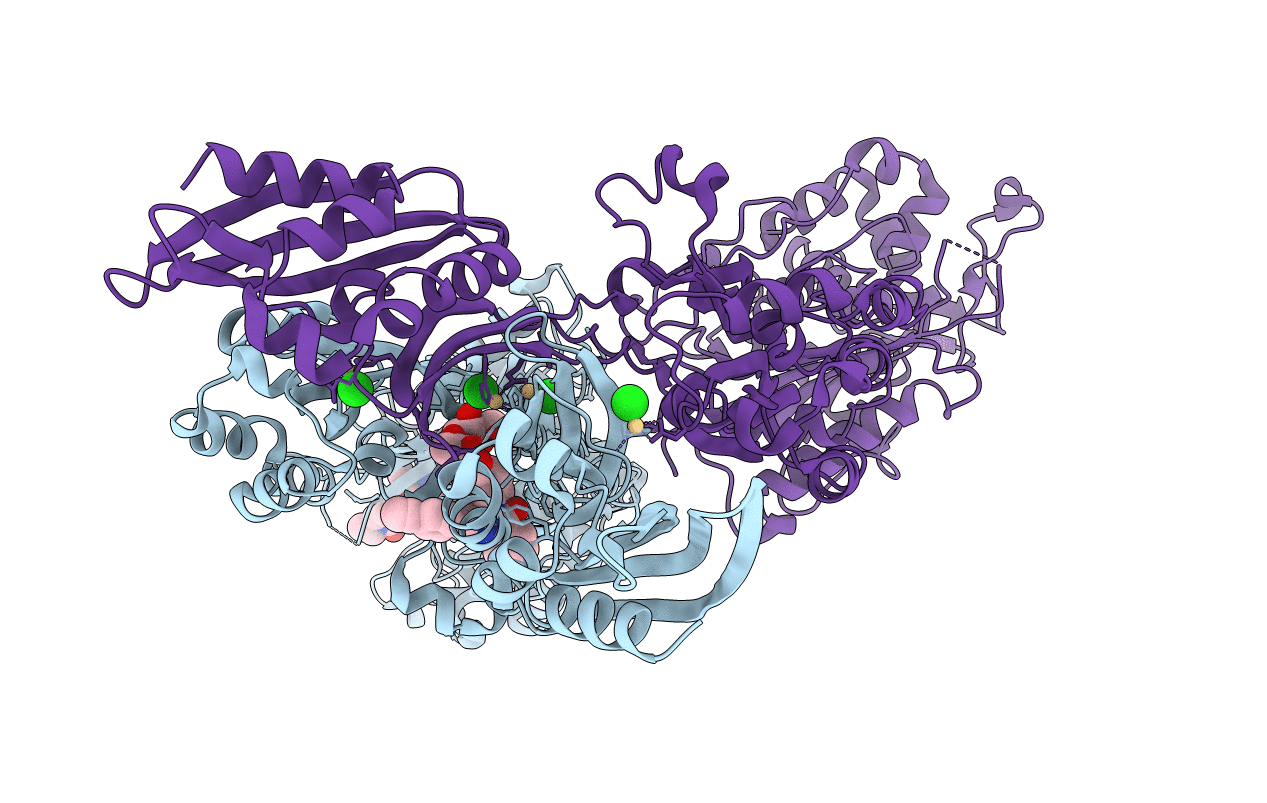 |
Crystal Structure Of Pbp2A From Mrsa In Complex With Piperacillin And Quinazolinone
Organism: Staphylococcus aureus (strain mu50 / atcc 700699)
Method: X-RAY DIFFRACTION Resolution:2.50 Å Release Date: 2019-11-27 Classification: HYDROLASE Ligands: QLN, JPP, CL, CD, MUR |
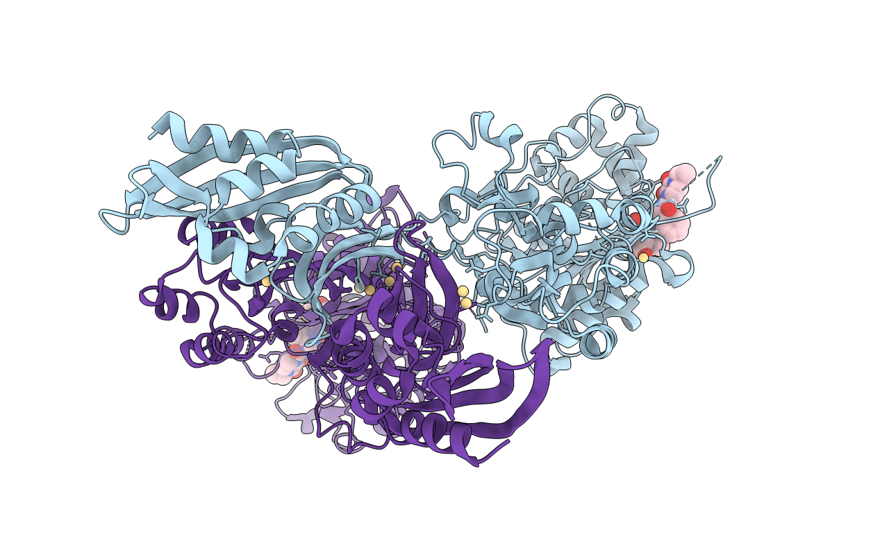 |
Crystal Structure Of Pbp2A From Mrsa In Complex With Piperacillin At Active Site.
Organism: Staphylococcus aureus (strain mu50 / atcc 700699)
Method: X-RAY DIFFRACTION Resolution:2.82 Å Release Date: 2019-08-14 Classification: HYDROLASE Ligands: JPP, CD |
 |
Organism: Homo sapiens
Method: X-RAY DIFFRACTION Resolution:2.00 Å Release Date: 2019-03-20 Classification: TRANSFERASE Ligands: NAG, CL, ZN |
 |
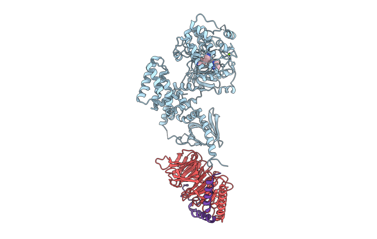
Organism: Homo sapiens, Bos taurus
Method: X-RAY DIFFRACTION Resolution:2.74 Å Release Date: 2018-04-25 Classification: TRANSFERASE/Signaling Protein Ligands: EJS |
 |
Organism: Homo sapiens, Bos taurus
Method: X-RAY DIFFRACTION Resolution:2.90 Å Release Date: 2017-12-27 Classification: Transferase/Signaling Protein Ligands: AFM, MG |
 |
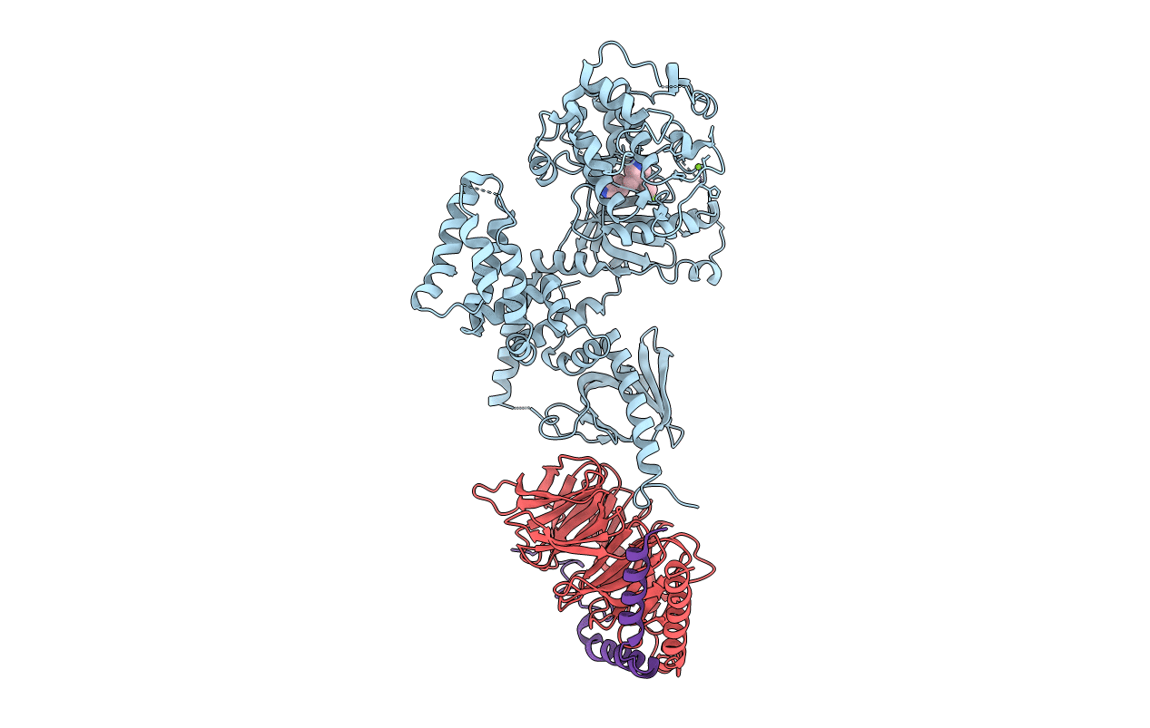
Organism: Homo sapiens, Bos taurus
Method: X-RAY DIFFRACTION Resolution:2.31 Å Release Date: 2017-12-27 Classification: TRANSFERASE/Signaling Protein Ligands: AFV |
 |
Organism: Homo sapiens, Bos taurus
Method: X-RAY DIFFRACTION Resolution:3.10 Å Release Date: 2017-12-27 Classification: TRANSFERASE Ligands: ZSO, MG |
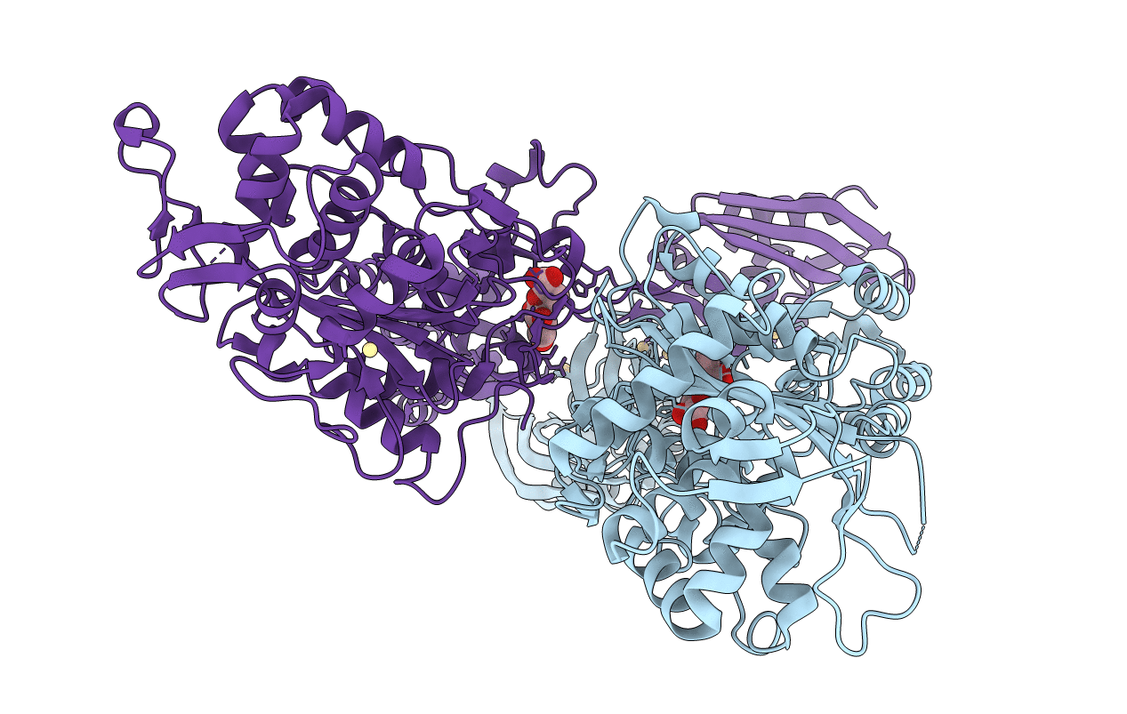 |
Organism: Staphylococcus aureus
Method: X-RAY DIFFRACTION Resolution:1.98 Å Release Date: 2017-02-08 Classification: Penicillin binding protein Ligands: CD, MUR |
 |
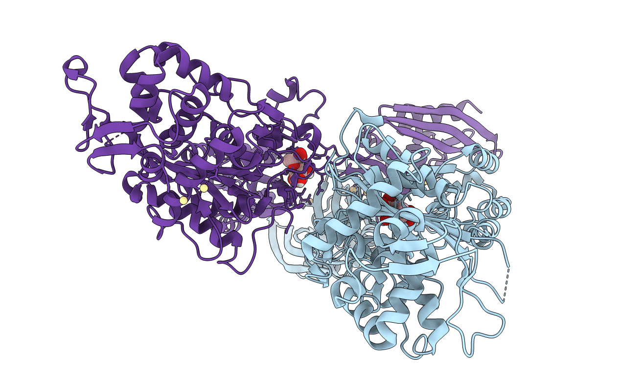
Organism: Staphylococcus aureus
Method: X-RAY DIFFRACTION Resolution:2.00 Å Release Date: 2017-02-08 Classification: Penicillin binding protein Ligands: CD, MUR |
 |
Organism: Staphylococcus aureus
Method: X-RAY DIFFRACTION Resolution:2.00 Å Release Date: 2017-02-08 Classification: Penicillin binding protein Ligands: CD, MUR |
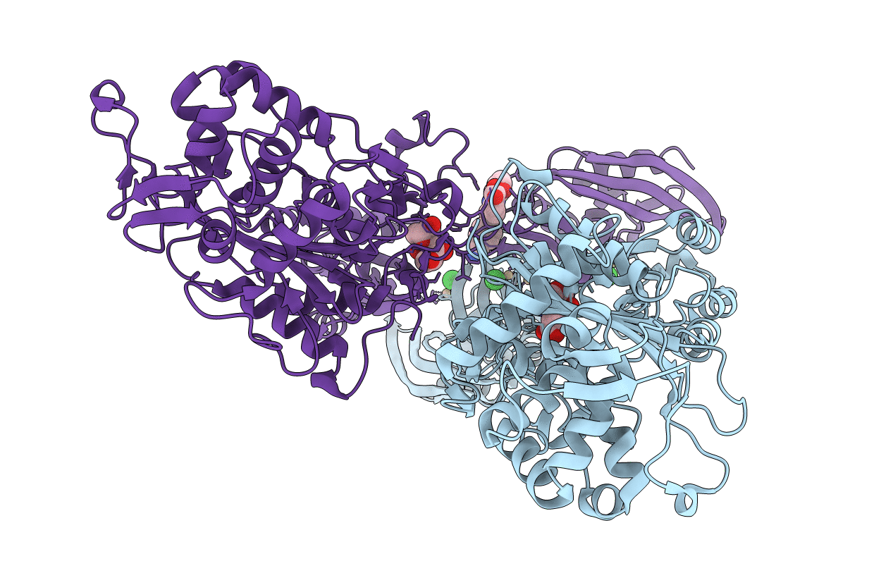 |
Organism: Staphylococcus aureus subsp. aureus mu50
Method: X-RAY DIFFRACTION Resolution:1.95 Å Release Date: 2015-02-11 Classification: HYDROLASE Ligands: CD, CL, MUR, QNZ |

