
Search Count: 54
 |
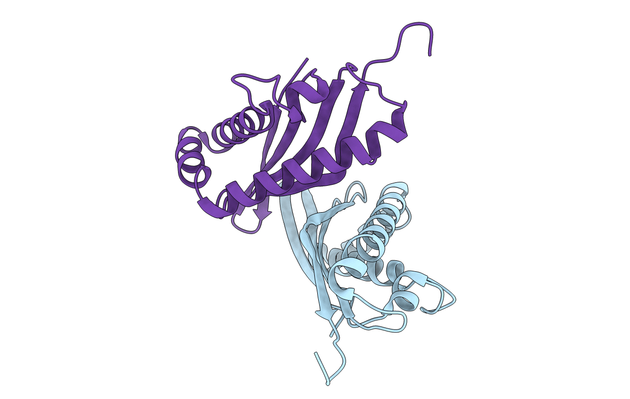
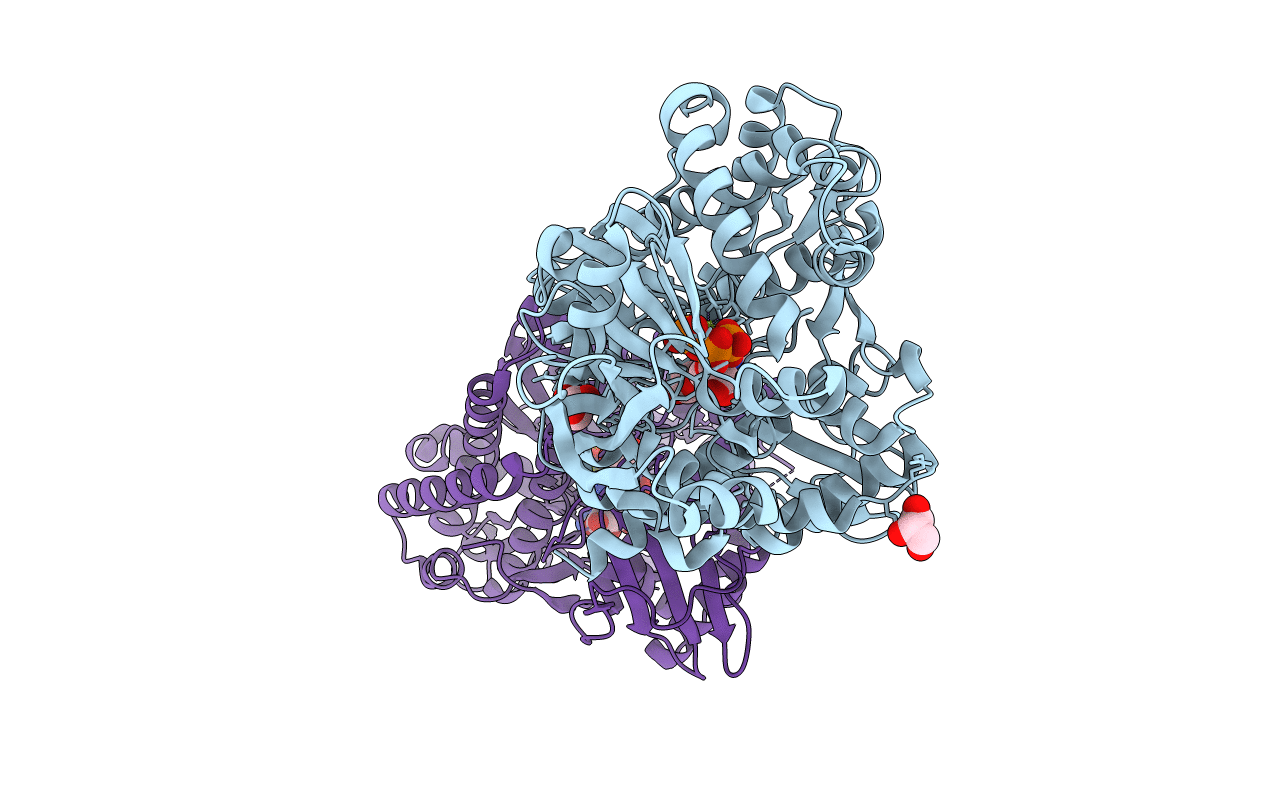
Organism: Gallus gallus, Rattus norvegicus, Sus scrofa
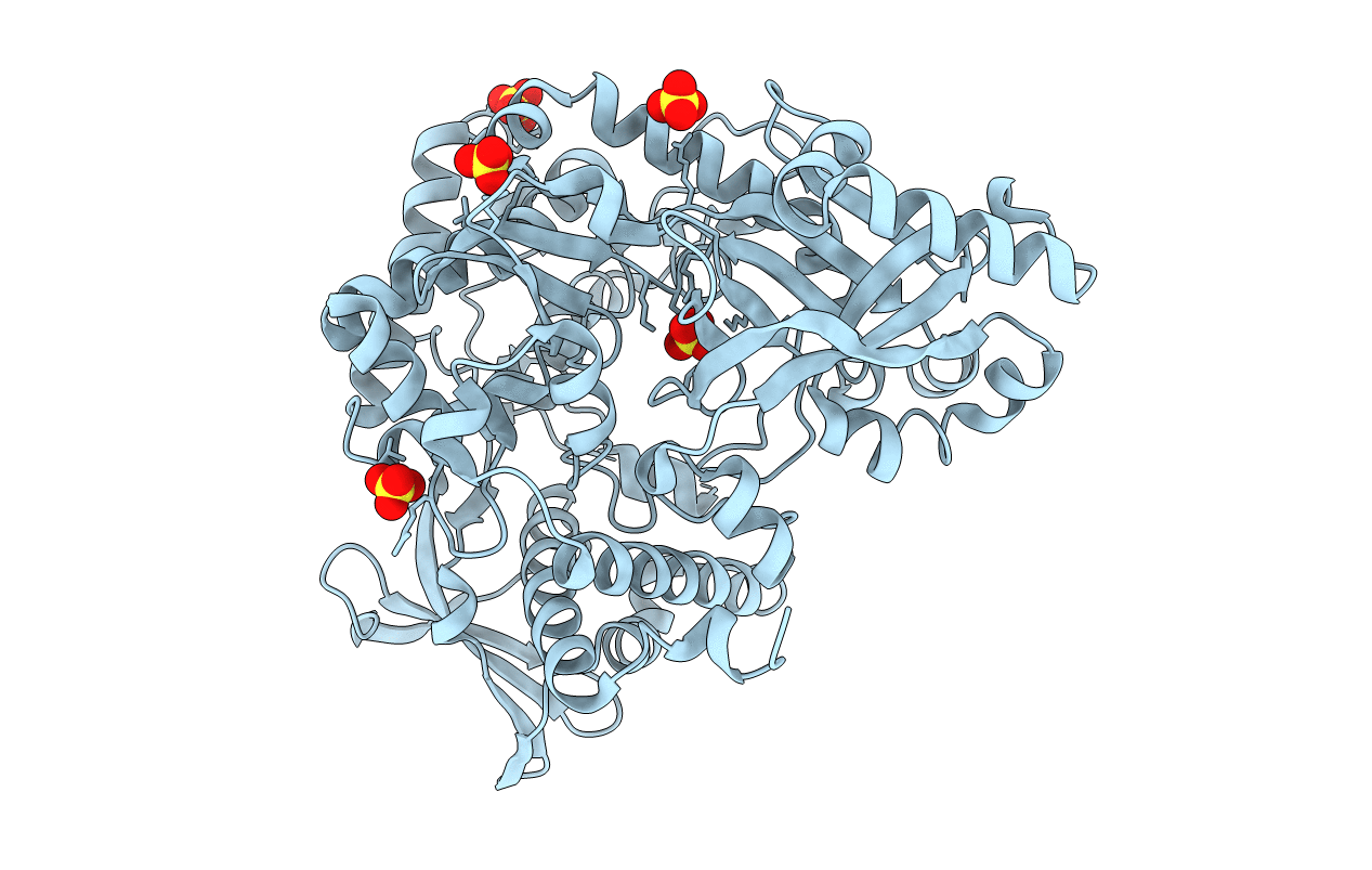
Method: X-RAY DIFFRACTION Resolution:2.61 Å Release Date: 2016-05-18 Classification: STRUCTURAL PROTEIN Ligands: GTP, MG, CA, GDP, MES, X3H, ACP |
 |
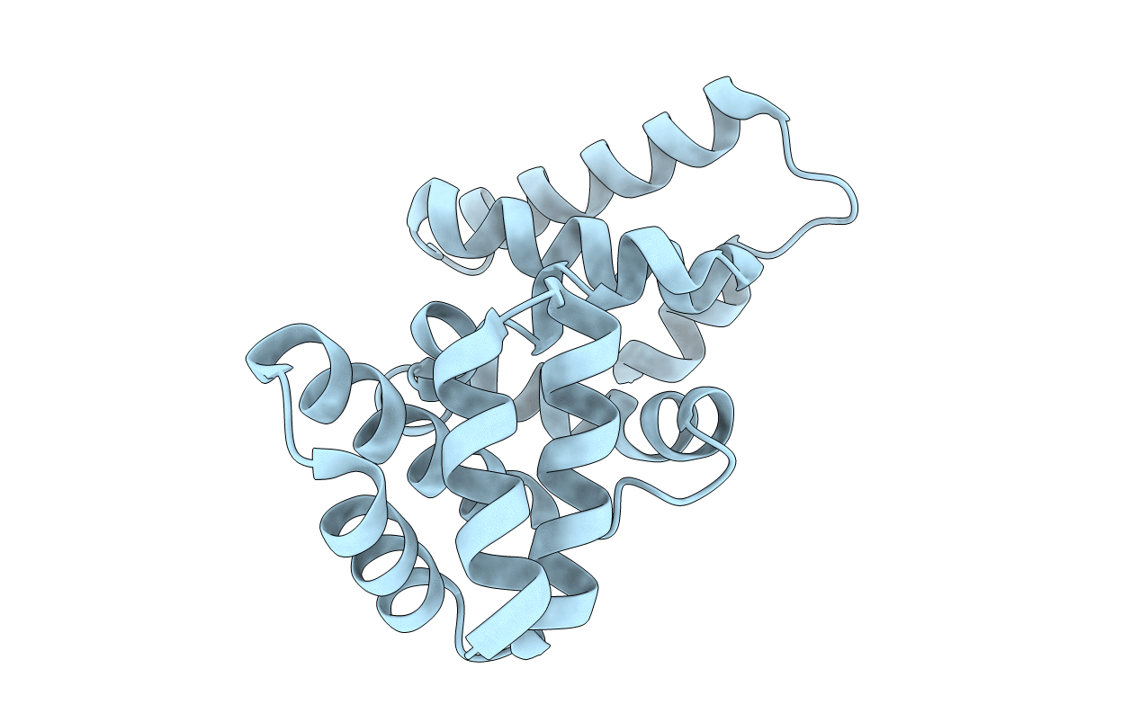
Organism: Rattus norvegicus, Gallus gallus, Sus scrofa
Method: X-RAY DIFFRACTION Resolution:2.24 Å Release Date: 2016-05-18 Classification: STRUCTURAL PROTEIN Ligands: GTP, MG, CA, CL, GDP, MES, ACP |
 |
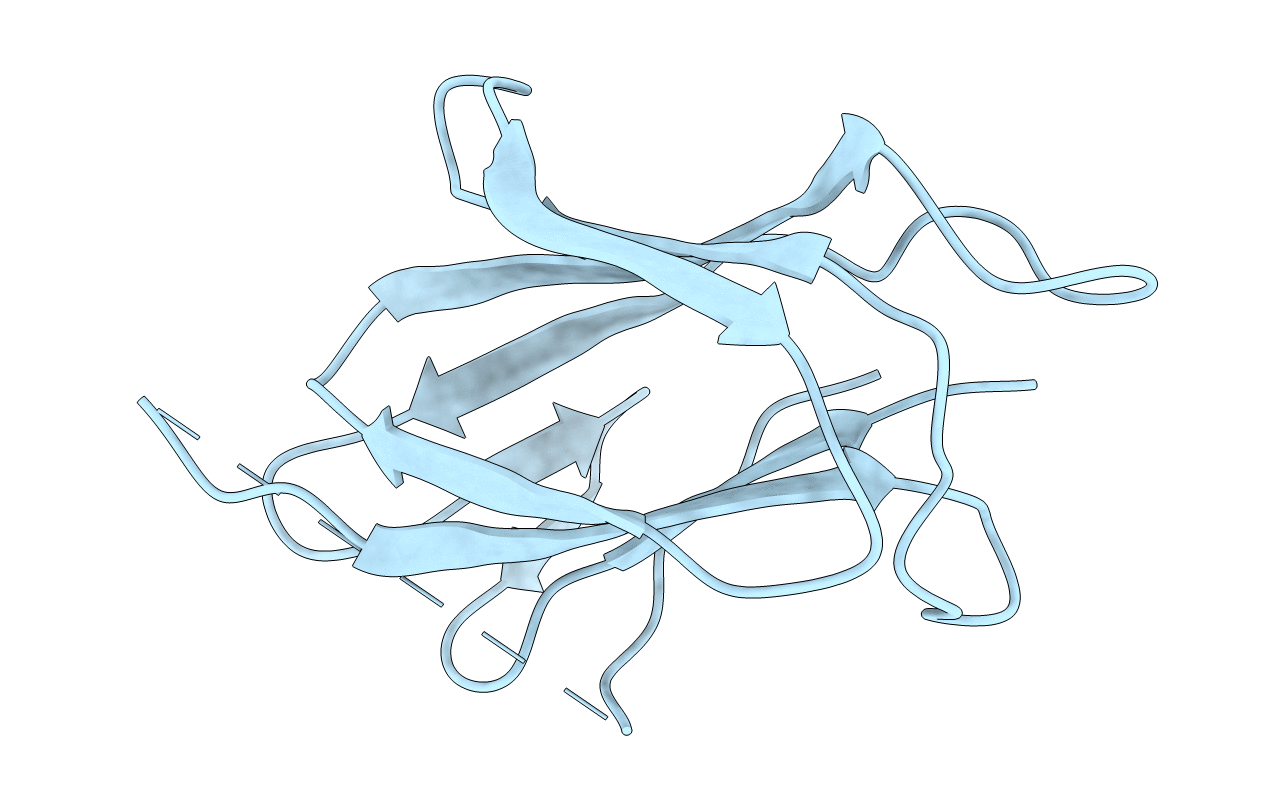
N-Terminal Thioester Domain Of Surface Protein From Clostridium Perfringens, Cys138Ala Mutant
Organism: Clostridium perfringens
Method: X-RAY DIFFRACTION Resolution:1.60 Å Release Date: 2015-06-03 Classification: CELL ADHESION Ligands: EDO |
 |
N-Terminal Thioester Domain Of Surface Protein From Clostridium Perfringens
Organism: Clostridium perfringens
Method: X-RAY DIFFRACTION Resolution:2.62 Å Release Date: 2015-06-03 Classification: CELL ADHESION |
 |
N-Terminal Thioester Domain Of Fibronectin-Binding Protein Sfbi From Streptococcus Pyogenes
Organism: Streptococcus pyogenes
Method: X-RAY DIFFRACTION Resolution:1.35 Å Release Date: 2015-06-03 Classification: CELL ADHESION Ligands: ZN, ACT |
 |
N-Terminal Thioester Domain Of Protein F2 Like Fibronectin-Binding Protein From Streptococcus Pneumoniae
Organism: Streptococcus pneumoniae
Method: X-RAY DIFFRACTION Resolution:1.30 Å Release Date: 2015-06-03 Classification: CELL ADHESION Ligands: MES, GOL |
 |
Crystal Structure Of Histidine-Rich Glycoprotein N2 Domain Reveals Redox Activity At An Interdomain Disulfide Bridge: Implications For The Regulation Of Angiogenesis
Organism: Oryctolagus cuniculus
Method: X-RAY DIFFRACTION Resolution:1.93 Å Release Date: 2014-02-19 Classification: BLOOD CLOTTING Ligands: GSH, GOL |
 |
Organism: Uncultured prochloron sp. 06037a
Method: X-RAY DIFFRACTION Resolution:2.90 Å Release Date: 2013-11-06 Classification: ISOMERASE Ligands: ZN |
 |
Organism: Prochloron didemni
Method: X-RAY DIFFRACTION Resolution:2.19 Å Release Date: 2012-07-18 Classification: HYDROLASE |
 |
Organism: Prochloron didemni, Synthetic construct
Method: X-RAY DIFFRACTION Resolution:2.63 Å Release Date: 2012-07-18 Classification: HYDROLASE/PEPTIDE |
 |
Thermodbp: A Non-Canonical Single-Stranded Dna Binding Protein With A Novel Structure And Mechanism
Organism: Thermoproteus tenax
Method: X-RAY DIFFRACTION Resolution:2.00 Å Release Date: 2011-11-23 Classification: DNA BINDING PROTEIN |
 |
Crystal Structure Of Hypothetical Protein Orf239 From Pyrobaculum Spherical Virus
Organism: Pyrobaculum spherical virus
Method: X-RAY DIFFRACTION Resolution:1.45 Å Release Date: 2011-02-16 Classification: UNKNOWN FUNCTION |
 |
Co-Complex Structure Of Achromobactin Synthetase Protein D (Acsd) With Atp And N-Citryl-Ethylenediamine From Pectobacterium Chrysanthemi
Organism: Erwinia chrysanthemi
Method: X-RAY DIFFRACTION Resolution:2.00 Å Release Date: 2011-01-19 Classification: LIGASE Ligands: ATP, MG, GOL, X3J |
 |
Crystal Structure Of The Streptococcus Pyogenes Fibronectin Binding Protein Fbab-B
Organism: Streptococcus pyogenes
Method: X-RAY DIFFRACTION Resolution:1.60 Å Release Date: 2010-09-01 Classification: PROTEIN BINDING |
 |
Apo Structure Of The Alcaligin Biosynthesis Protein C (Alcc) From Bordetella Bronchiseptica
Organism: Bordetella bronchiseptica
Method: X-RAY DIFFRACTION Resolution:2.40 Å Release Date: 2010-07-28 Classification: BIOSYNTHETIC PROTEIN Ligands: SO4 |
 |
Organism: Sulfolobus solfataricus
Method: X-RAY DIFFRACTION Resolution:2.70 Å Release Date: 2010-07-28 Classification: UNKNOWN FUNCTION |
 |
Crystal Structure Of 3-Oxoacyl-(Acyl Carrier Protein) Synthase Iii, Fabh From Pseudomonas Aeruginosa Pao1
Organism: Pseudomonas aeruginosa
Method: X-RAY DIFFRACTION Resolution:1.81 Å Release Date: 2010-07-28 Classification: TRANSFERASE |
 |
Crystal Structure Of Hypothetical Protein Sso1986 From Sulfolobus Solfataricus P2
Organism: Sulfolobus solfataricus
Method: X-RAY DIFFRACTION Resolution:2.05 Å Release Date: 2010-07-28 Classification: UNKNOWN FUNCTION |
 |
Crystal Structure Of The Hypothetical Protein Orf126 From Pyrobaculum Spherical Virus
Organism: Pyrobaculum spherical virus
Method: X-RAY DIFFRACTION Resolution:2.00 Å Release Date: 2010-07-28 Classification: VIRAL PROTEIN Ligands: ZN |
 |
Organism: Sulfolobus islandicus rudivirus 1
Method: X-RAY DIFFRACTION Resolution:2.20 Å Release Date: 2010-07-28 Classification: VIRAL PROTEIN Ligands: MLI |