
Search Count: 11
 |
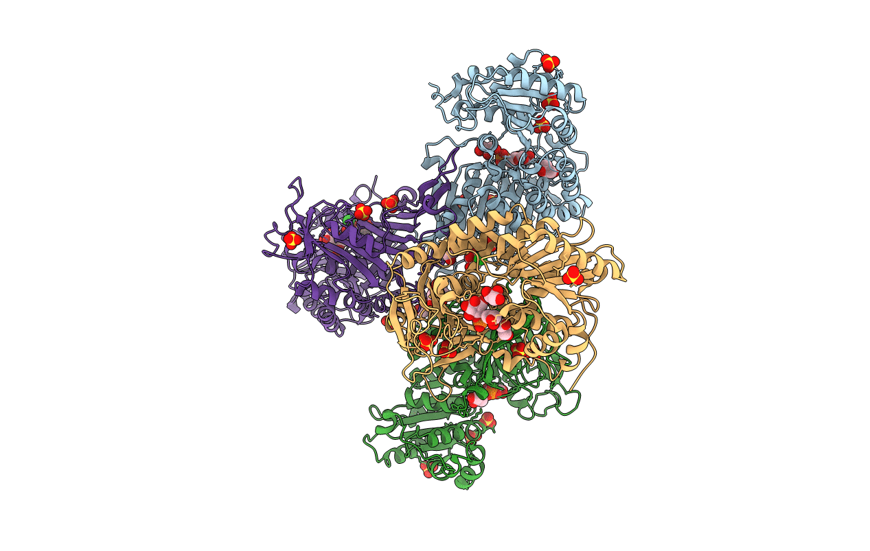
Organism: Trypanosoma cruzi
Method: X-RAY DIFFRACTION Resolution:2.85 Å Release Date: 2018-05-02 Classification: OXIDOREDUCTASE Ligands: SO4, GOL, CL |
 |
Organism: Trypanosoma cruzi
Method: X-RAY DIFFRACTION Resolution:3.35 Å Release Date: 2018-05-02 Classification: OXIDOREDUCTASE Ligands: BG6, SO4, GOL, CL |
 |
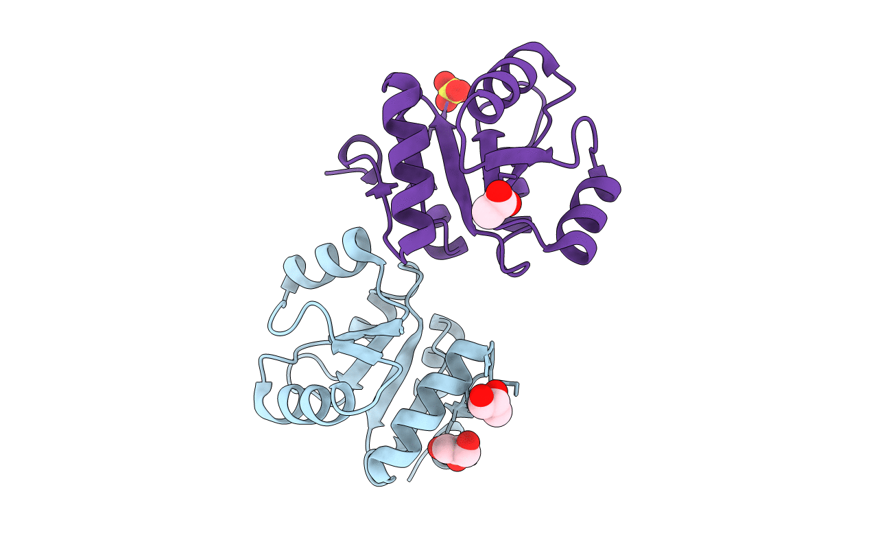
Crystal Structure Of The Full-Length Response Regulator Desr In The Active State
Organism: Bacillus subtilis subsp. subtilis
Method: X-RAY DIFFRACTION Resolution:2.31 Å Release Date: 2014-07-16 Classification: DNA BINDING PROTEIN Ligands: MG, BEF, GOL, NA |
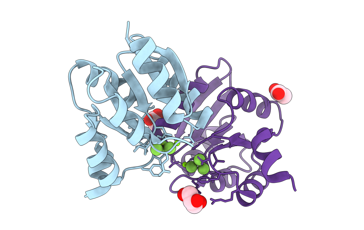 |
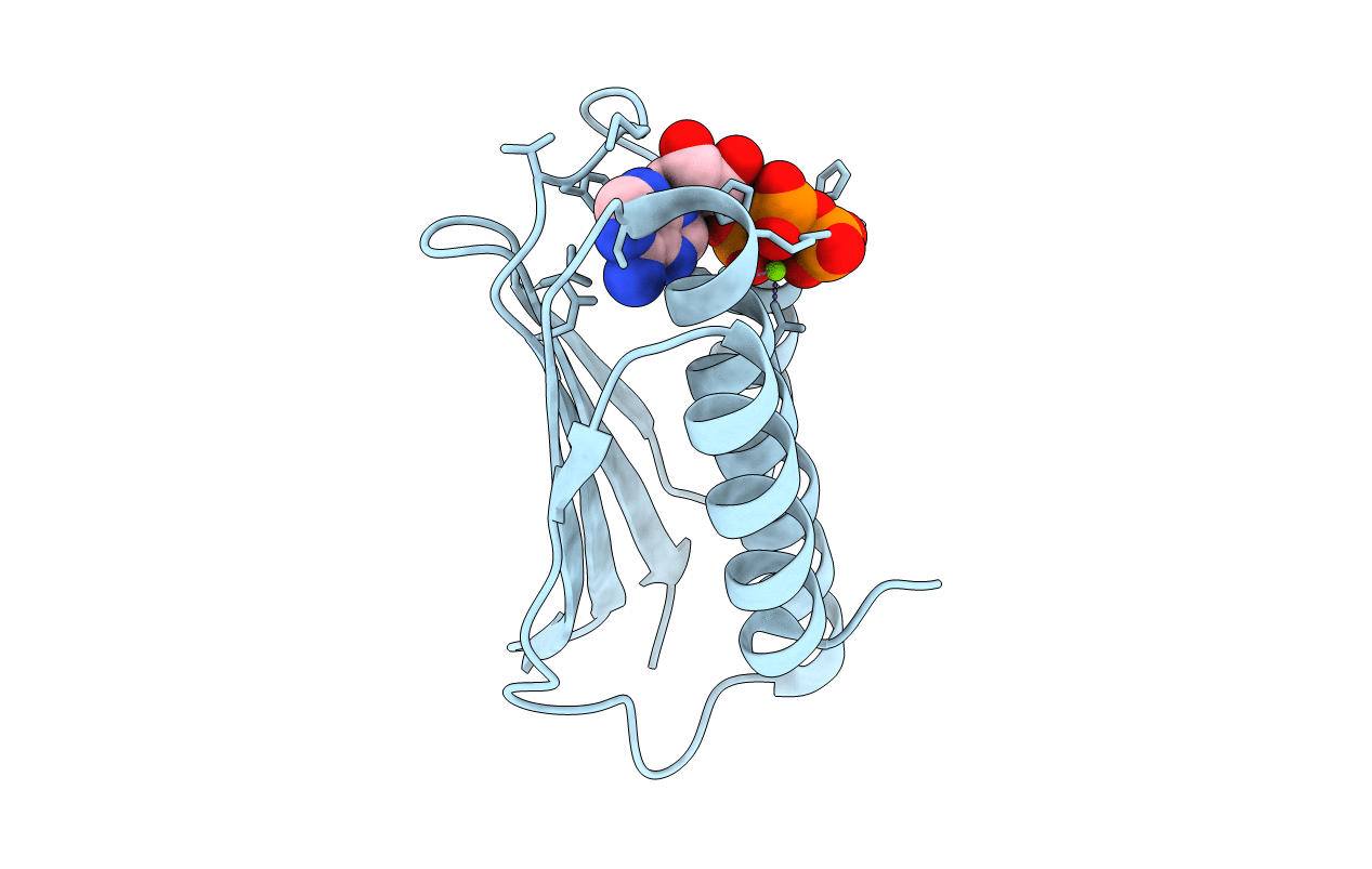
Crystal Structure Of The Receiver Domain Of Desr In Complex With Beryllofluoride And Magnesium
Organism: Bacillus subtilis subsp. subtilis
Method: X-RAY DIFFRACTION Resolution:2.27 Å Release Date: 2014-07-16 Classification: DNA BINDING PROTEIN Ligands: MG, BEF, ACT, GOL |
 |
Organism: Bacillus subtilis subsp. subtilis
Method: X-RAY DIFFRACTION Resolution:1.95 Å Release Date: 2014-07-16 Classification: DNA BINDING PROTEIN Ligands: GOL, SO4, ACT |
 |
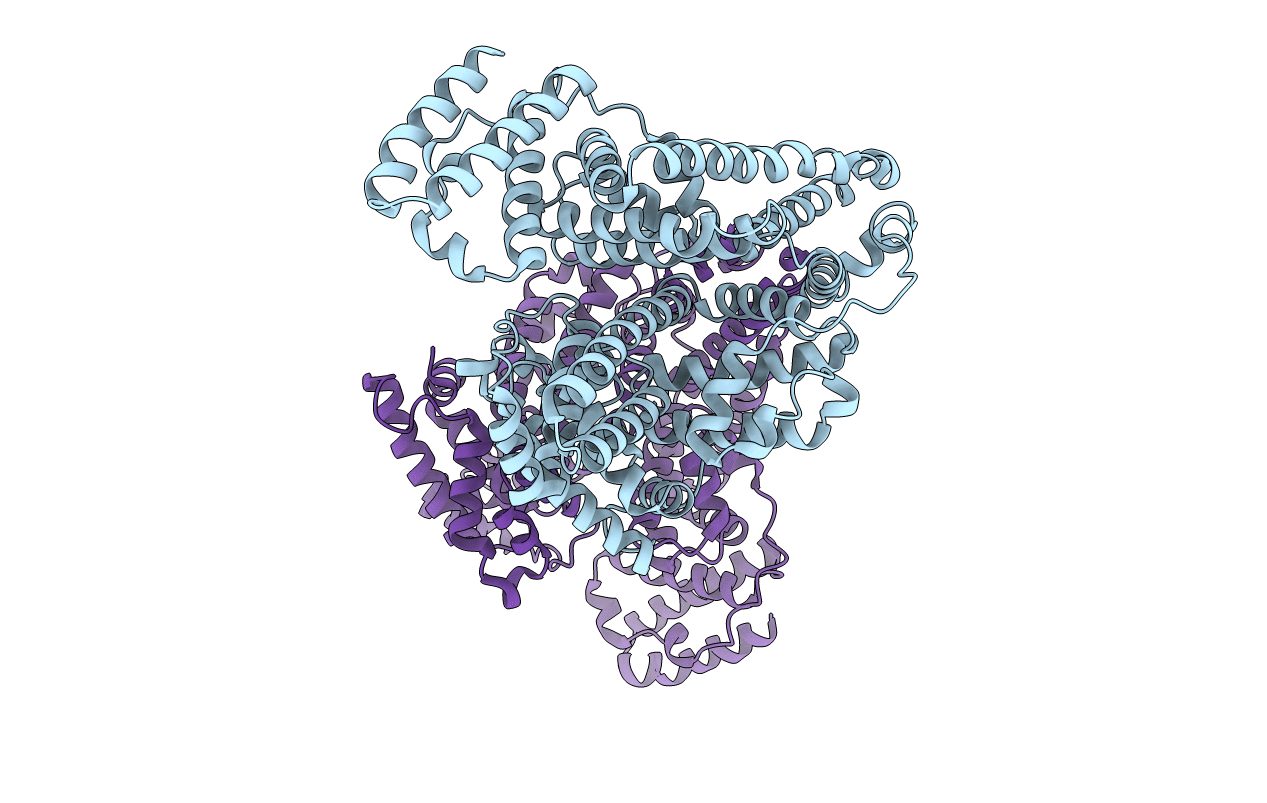
Crystal Structure Of The Unphosphorylated Receiver Domain Of Desr In The Active State
Organism: Bacillus subtilis subsp. subtilis
Method: X-RAY DIFFRACTION Resolution:2.54 Å Release Date: 2014-07-16 Classification: DNA BINDING PROTEIN Ligands: PO4, K |
 |
Organism: Leptospira biflexa serovar patoc
Method: X-RAY DIFFRACTION Resolution:1.44 Å Release Date: 2014-05-14 Classification: SIGNALING PROTEIN Ligands: GOL, SO4 |
 |
Crystal Structure Analysis Of Human Serum Albumin In Complex With Chloride Anions At Cryogenic Temperature
Organism: Homo sapiens
Method: X-RAY DIFFRACTION Resolution:2.30 Å Release Date: 2012-04-25 Classification: PROTEIN BINDING Ligands: CL |
 |
Crystal Structure Of An Engineered Ferredoxin Nadp Reductase (Fnr) From Pisum Sativum
Organism: Pisum sativum
Method: X-RAY DIFFRACTION Resolution:2.90 Å Release Date: 2011-02-23 Classification: OXIDOREDUCTASE Ligands: FAD, SO4 |
 |
Crystal Structure Of An Engineered Ferredoxin(Flavodoxin) Nadp(H) Reductase (Fpr) From Escherichia Coli
Organism: Escherichia coli
Method: X-RAY DIFFRACTION Resolution:1.90 Å Release Date: 2011-02-23 Classification: OXIDOREDUCTASE Ligands: FAD, NAP, TRS, GOL, ZN, NA |
 |
Organism: Bacillus subtilis
Method: X-RAY DIFFRACTION Resolution:1.74 Å Release Date: 2009-09-15 Classification: TRANSFERASE Ligands: ATP, MG, IOD |