
Search Count: 65
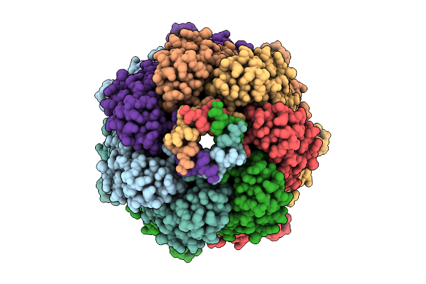 |
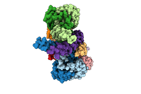
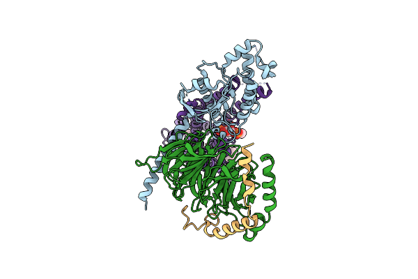
Ynai In Its Open Conformation Purified In Ddm Showing Ligand-Filled Pockets
Organism: Escherichia coli
Method: ELECTRON MICROSCOPY Release Date: 2025-09-24 Classification: MEMBRANE PROTEIN Ligands: D12 |
 |
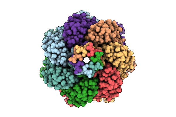
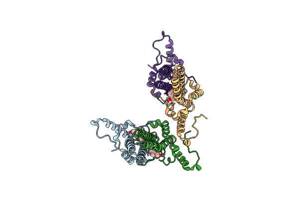
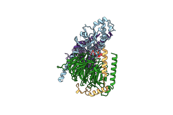
A Ynai-Mscs Chimera In A Closed Conformation Purified In Ddm With Additional Lipids Showing Ligand-Filled Pore And Pockets
Organism: Escherichia coli (strain k12)
Method: ELECTRON MICROSCOPY Release Date: 2025-09-24 Classification: MEMBRANE PROTEIN Ligands: D12 |
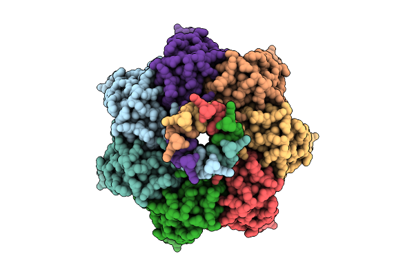 |
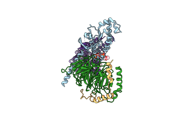
A Ynai-Mscs Chimera In An Open Conformation Purified In Ddm Showing Ligand-Filled Pockets
Organism: Escherichia coli (strain k12)
Method: ELECTRON MICROSCOPY Release Date: 2025-09-24 Classification: MEMBRANE PROTEIN Ligands: D12 |
 |
Ynai In Closed Conformation Purified In Ddm With Additional Lipids Showing Ligand-Filled Pockets
Organism: Escherichia coli
Method: ELECTRON MICROSCOPY Release Date: 2025-09-24 Classification: MEMBRANE PROTEIN Ligands: D12 |
 |
Organism: Cricetulus griseus
Method: ELECTRON MICROSCOPY Release Date: 2025-09-17 Classification: NUCLEAR PROTEIN |
 |
Organism: Cricetulus griseus
Method: ELECTRON MICROSCOPY Release Date: 2025-09-10 Classification: NUCLEAR PROTEIN |
 |
Organism: Cricetulus griseus
Method: ELECTRON MICROSCOPY Release Date: 2025-09-10 Classification: NUCLEAR PROTEIN |
 |
Organism: Escherichia coli o127:h6
Method: ELECTRON MICROSCOPY Release Date: 2025-07-16 Classification: TOXIN Ligands: MN |
 |
Organism: Escherichia coli o127:h6
Method: ELECTRON MICROSCOPY Release Date: 2025-07-16 Classification: TOXIN |
 |
Organism: Escherichia coli o127:h6
Method: ELECTRON MICROSCOPY Release Date: 2025-07-16 Classification: TOXIN |
 |
Organism: Homo sapiens
Method: ELECTRON MICROSCOPY Release Date: 2025-07-02 Classification: MEMBRANE PROTEIN Ligands: CLR, T7M, ZKV |
 |
Organism: Escherichia coli o127:h6 str. e2348/69
Method: ELECTRON MICROSCOPY Release Date: 2025-04-09 Classification: TOXIN |
 |
Organism: Escherichia coli o127:h6 str. e2348/69
Method: ELECTRON MICROSCOPY Release Date: 2025-04-09 Classification: TOXIN |
 |
Organism: Hepatitis b virus ayw/france/tiollais/1979
Method: ELECTRON MICROSCOPY Release Date: 2024-07-31 Classification: VIRUS LIKE PARTICLE Ligands: 64Z |
 |
Organism: Hepatitis b virus, Synthetic construct
Method: ELECTRON MICROSCOPY Release Date: 2024-07-31 Classification: VIRUS LIKE PARTICLE |
 |
Organism: Hepatitis b virus, Synthetic construct
Method: ELECTRON MICROSCOPY Release Date: 2024-07-31 Classification: VIRUS LIKE PARTICLE |
 |
Organism: Homo sapiens
Method: ELECTRON MICROSCOPY Release Date: 2024-05-29 Classification: MEMBRANE PROTEIN Ligands: CLR, ZL9, T7M |
 |
Organism: Homo sapiens
Method: ELECTRON MICROSCOPY Release Date: 2024-05-29 Classification: MEMBRANE PROTEIN Ligands: CLR, T7M, ZKV |
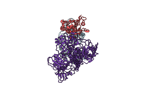 |
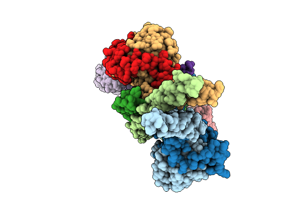
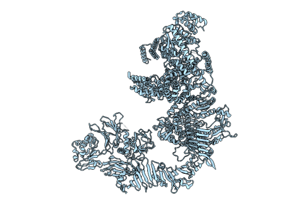
Cryo-Em Structure Of The Dna Polymerase Holoenzyme E9-A20-D4 Of Vaccinia Virus
Organism: Vaccinia virus copenhagen
Method: ELECTRON MICROSCOPY Release Date: 2024-05-08 Classification: VIRAL PROTEIN |
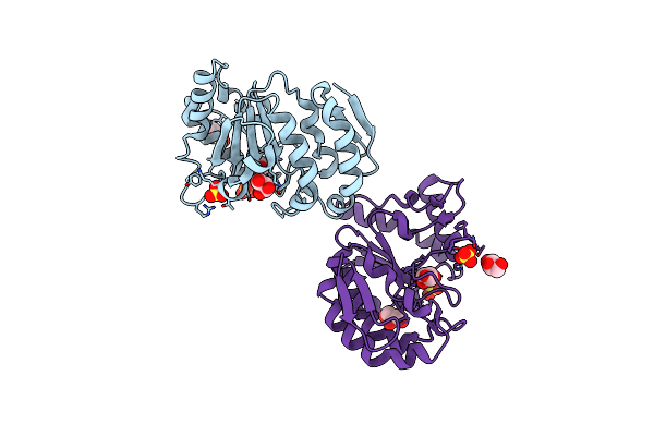 |
Organism: Vaccinia virus copenhagen
Method: X-RAY DIFFRACTION Resolution:1.32 Å Release Date: 2024-05-08 Classification: HYDROLASE Ligands: GOL, SO4 |