
Search Count: 17
 |
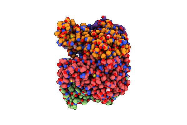
Organism: Homo sapiens
Method: X-RAY DIFFRACTION Resolution:2.71 Å Release Date: 2024-01-31 Classification: TRANSFERASE |
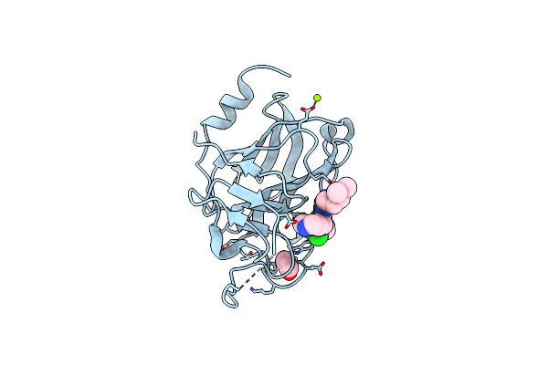 |
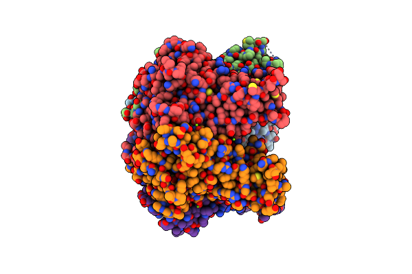
Organism: Homo sapiens
Method: X-RAY DIFFRACTION Resolution:1.30 Å Release Date: 2024-01-31 Classification: TRANSFERASE Ligands: YCB, MG, CL, EDO |
 |
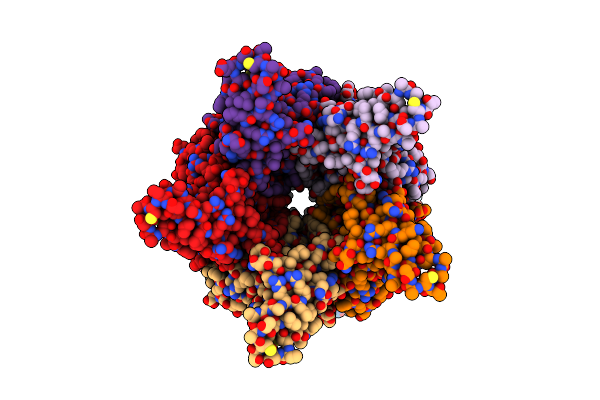
Organism: Saccharomyces cerevisiae
Method: ELECTRON MICROSCOPY Release Date: 2024-01-17 Classification: LIGASE Ligands: PCW, PX6 |
 |
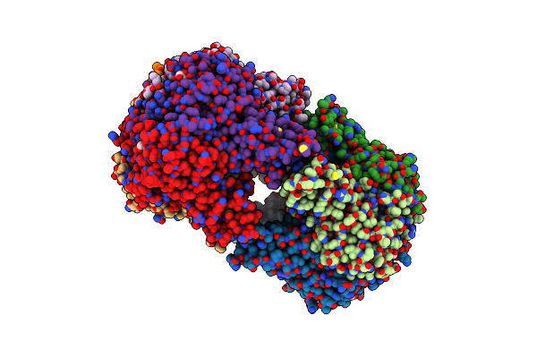
Organism: Saccharomyces cerevisiae
Method: ELECTRON MICROSCOPY Release Date: 2024-01-17 Classification: LIGASE Ligands: PCW, PX6 |
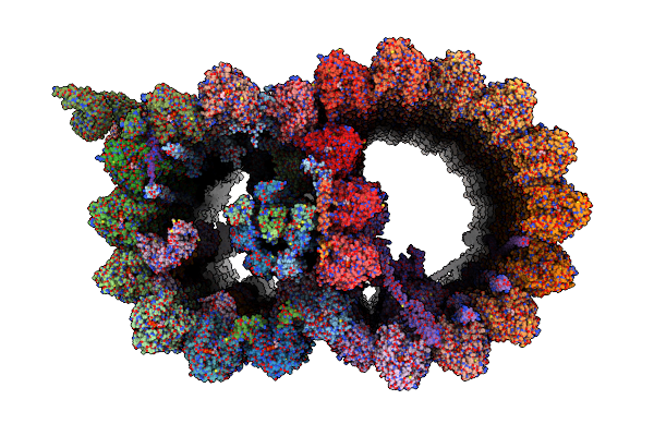 |
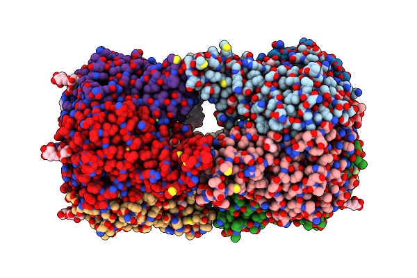
Structure Of The 48-Nm Repeat Doublet Microtubule From Bovine Tracheal Cilia
Organism: Bos taurus
Method: ELECTRON MICROSCOPY Release Date: 2021-10-27 Classification: STRUCTURAL PROTEIN Ligands: GTP, MG, GDP |
 |
Organism: New jersey polyomavirus-2013
Method: X-RAY DIFFRACTION Resolution:1.80 Å Release Date: 2020-07-22 Classification: VIRAL PROTEIN Ligands: GOL, MG |
 |
Organism: New jersey polyomavirus-2013
Method: X-RAY DIFFRACTION Resolution:2.30 Å Release Date: 2020-07-08 Classification: VIRAL PROTEIN Ligands: GOL, MG |
 |
Organism: Human polyomavirus 12
Method: X-RAY DIFFRACTION Resolution:1.55 Å Release Date: 2020-07-08 Classification: VIRAL PROTEIN Ligands: GOL |
 |
Structure Of Human Polyomavirus 12 Vp1 In Complex With 3'-Sialyllactosamine
Organism: Human polyomavirus 12
Method: X-RAY DIFFRACTION Resolution:1.80 Å Release Date: 2020-07-08 Classification: VIRAL PROTEIN Ligands: PO4, SIA |
 |
Organism: Sheep polyomavirus 1
Method: X-RAY DIFFRACTION Resolution:2.45 Å Release Date: 2020-07-08 Classification: VIRAL PROTEIN Ligands: GOL |
 |
Organism: Sheep polyomavirus 1
Method: X-RAY DIFFRACTION Resolution:1.65 Å Release Date: 2020-07-08 Classification: VIRAL PROTEIN Ligands: MPD, MG, SIA |
 |
Organism: Sheep polyomavirus 1
Method: X-RAY DIFFRACTION Resolution:1.60 Å Release Date: 2020-07-08 Classification: VIRAL PROTEIN Ligands: GOL, EDO, MG, SIA |
 |
Organism: Goose hemorrhagic polyomavirus
Method: X-RAY DIFFRACTION Resolution:1.45 Å Release Date: 2020-07-08 Classification: VIRAL PROTEIN Ligands: IMD, EDO, CL |
 |
Structure Of Goose Hemorrhagic Polyomavirus Vp1 In Complex With 2-O-Methyl-5-N-Acetyl-Alpha-D-Neuraminic Acid
Organism: Goose hemorrhagic polyomavirus
Method: X-RAY DIFFRACTION Resolution:1.95 Å Release Date: 2020-07-08 Classification: VIRAL PROTEIN Ligands: MNA, IMD, EDO |
 |
Organism: Finch polyomavirus
Method: X-RAY DIFFRACTION Resolution:2.62 Å Release Date: 2020-07-08 Classification: VIRAL PROTEIN Ligands: CL |
 |
Structure Of Finch Polyomavirus Vp1 In Complex With 2-O-Methyl-5-N-Acetyl-Alpha-D-Neuraminic Acid
Organism: Finch polyomavirus
Method: X-RAY DIFFRACTION Resolution:2.65 Å Release Date: 2020-07-08 Classification: VIRAL PROTEIN Ligands: SIA, CL, MG |
 |
Organism: Chimpanzee polyomavirus bob
Method: X-RAY DIFFRACTION Resolution:1.90 Å Release Date: 2020-07-08 Classification: VIRAL PROTEIN Ligands: MG |