Search Count: 32
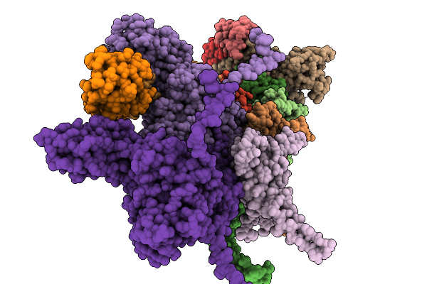 |
Organism: Escherichia phage johannrwettstein
Method: ELECTRON MICROSCOPY Release Date: 2025-11-19 Classification: VIRAL PROTEIN |
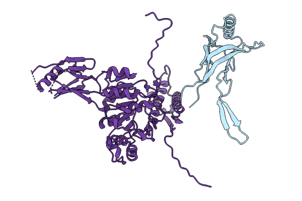 |
Organism: Escherichia phage johannrwettstein
Method: ELECTRON MICROSCOPY Release Date: 2025-11-19 Classification: VIRAL PROTEIN |
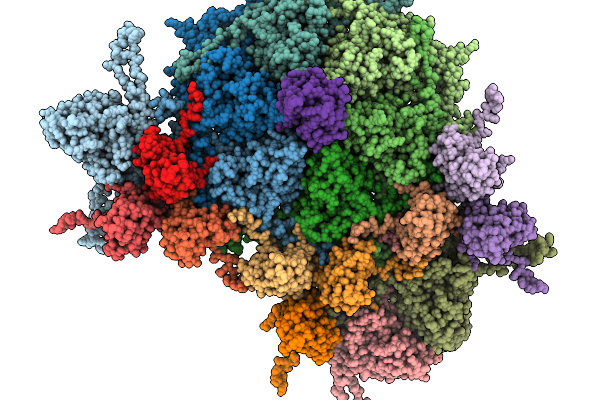 |
Organism: Escherichia phage johannrwettstein
Method: ELECTRON MICROSCOPY Release Date: 2025-11-19 Classification: VIRAL PROTEIN |
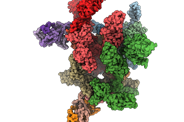 |
Organism: Escherichia phage johannrwettstein
Method: ELECTRON MICROSCOPY Release Date: 2025-11-19 Classification: VIRAL PROTEIN |
 |
Organism: Seneca valley virus usa/ssv-001
Method: ELECTRON MICROSCOPY Release Date: 2025-08-20 Classification: VIRUS Ligands: CA |
 |
Seneca Valley Virus Altered Particle At Physiological Condition (A-Particle[P])
Organism: Seneca valley virus usa/ssv-001
Method: ELECTRON MICROSCOPY Release Date: 2025-08-20 Classification: VIRUS Ligands: CA |
 |
Seneca Valley Virus Empty Rotated Particle At Acidic Condition (Er-Particle[C])
Organism: Seneca valley virus usa/ssv-001
Method: ELECTRON MICROSCOPY Release Date: 2025-08-20 Classification: VIRUS Ligands: CA |
 |
Seneca Valley Virus Empty Rotated Particle At Physiological Condition (Er-Particle[P])
Organism: Seneca valley virus usa/ssv-001
Method: ELECTRON MICROSCOPY Release Date: 2025-08-20 Classification: VIRUS Ligands: CA |
 |
Organism: Pectobacterium phage phite
Method: ELECTRON MICROSCOPY Release Date: 2025-04-16 Classification: VIRAL PROTEIN |
 |
Organism: Pectobacterium phage phite
Method: ELECTRON MICROSCOPY Release Date: 2025-04-16 Classification: VIRAL PROTEIN |
 |
Organism: Pectobacterium phage phite
Method: ELECTRON MICROSCOPY Release Date: 2025-04-16 Classification: VIRAL PROTEIN |
 |
Organism: Pectobacterium phage phite
Method: ELECTRON MICROSCOPY Release Date: 2025-04-16 Classification: VIRAL PROTEIN |
 |
Organism: Pectobacterium phage phite
Method: ELECTRON MICROSCOPY Release Date: 2025-04-16 Classification: VIRAL PROTEIN |
 |
Organism: Pectobacterium phage phite
Method: ELECTRON MICROSCOPY Release Date: 2025-04-16 Classification: VIRUS |
 |
Organism: Escherichia phage n4
Method: ELECTRON MICROSCOPY Release Date: 2025-02-19 Classification: VIRAL PROTEIN |
 |
Organism: Pectobacterium phage phim1
Method: ELECTRON MICROSCOPY Release Date: 2024-10-23 Classification: VIRUS |
 |
Organism: Pectobacterium phage phim1
Method: ELECTRON MICROSCOPY Release Date: 2024-10-23 Classification: VIRAL PROTEIN |
 |
Organism: Pectobacterium phage phim1
Method: ELECTRON MICROSCOPY Release Date: 2024-10-23 Classification: VIRAL PROTEIN |
 |
Organism: Pectobacterium phage phim1
Method: ELECTRON MICROSCOPY Release Date: 2024-10-23 Classification: VIRAL PROTEIN |
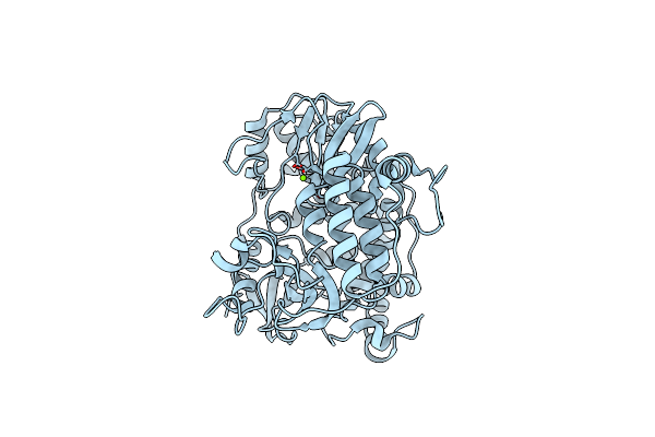 |
Organism: Norovirus hu/gii.4/sydney/nsw0514/2012/au
Method: ELECTRON MICROSCOPY Release Date: 2024-10-16 Classification: VIRAL PROTEIN Ligands: MG |

