
Search Count: 9
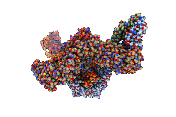 |
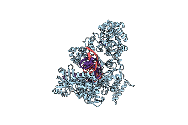
Organism: Homo sapiens, Escherichia coli (strain k12)
Method: ELECTRON MICROSCOPY Release Date: 2025-07-02 Classification: DNA BINDING PROTEIN |
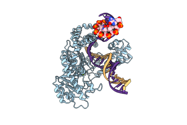 |
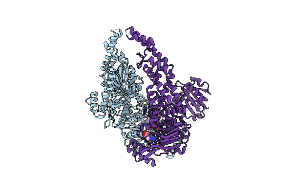
Organism: Escherichia coli 'bl21-gold(de3)plyss ag', Synthetic construct
Method: ELECTRON MICROSCOPY Release Date: 2023-08-09 Classification: DNA BINDING PROTEIN Ligands: MG |
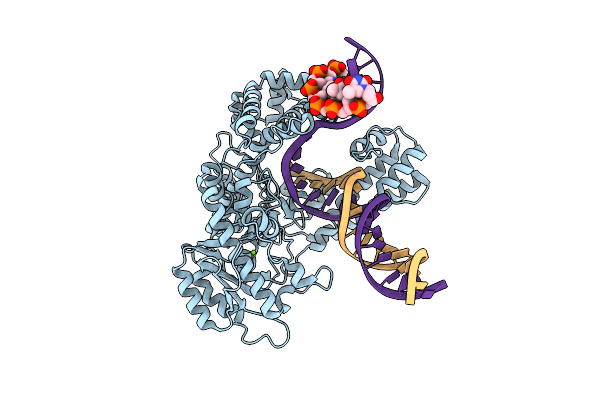 |
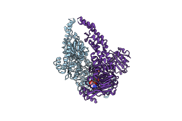
Organism: Escherichia coli 'bl21-gold(de3)plyss ag', Synthetic construct
Method: ELECTRON MICROSCOPY Release Date: 2023-08-09 Classification: DNA BINDING PROTEIN Ligands: MG |
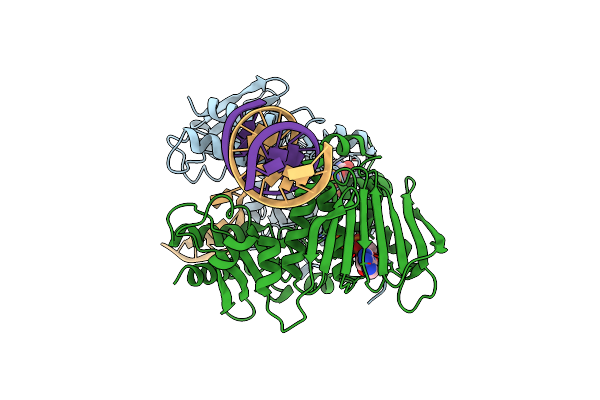 |
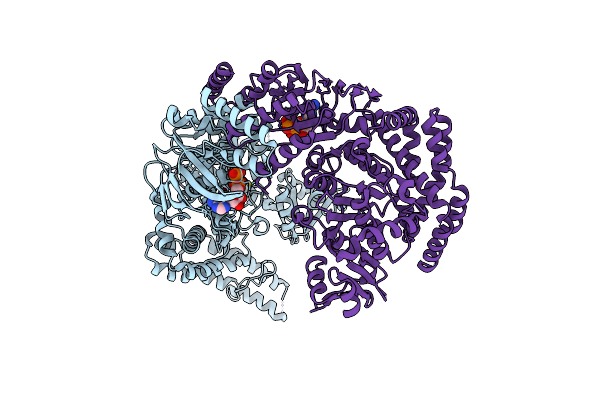
Organism: Escherichia coli (strain k12), Dna molecule
Method: ELECTRON MICROSCOPY Resolution:3.60 Å Release Date: 2022-06-29 Classification: DNA BINDING PROTEIN Ligands: ANP, MG |
 |
Organism: Mycobacterium tuberculosis, Synthetic construct
Method: ELECTRON MICROSCOPY Release Date: 2022-02-23 Classification: REPLICATION Ligands: ZN, 82W |
 |
Organism: Escherichia coli (strain k12)
Method: ELECTRON MICROSCOPY Release Date: 2022-01-12 Classification: DNA BINDING PROTEIN Ligands: MG, ANP |
 |
Organism: Escherichia coli (strain k12)
Method: ELECTRON MICROSCOPY Release Date: 2022-01-12 Classification: DNA BINDING PROTEIN Ligands: MG, ADP, VO4 |
 |
Organism: Escherichia coli
Method: ELECTRON MICROSCOPY Release Date: 2022-01-12 Classification: DNA BINDING PROTEIN Ligands: ADP |
 |
Organism: Escherichia coli
Method: ELECTRON MICROSCOPY Resolution:3.30 Å Release Date: 2022-01-12 Classification: DNA BINDING PROTEIN Ligands: MG, ATP, ADP |