
Search Count: 34
 |
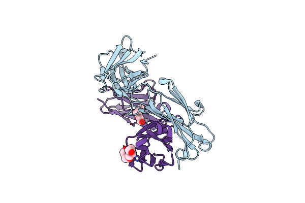
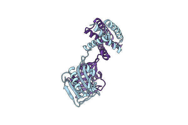
Organism: Homo sapiens
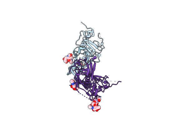
Method: X-RAY DIFFRACTION Resolution:2.60 Å Release Date: 2021-12-01 Classification: IMMUNE SYSTEM |
 |
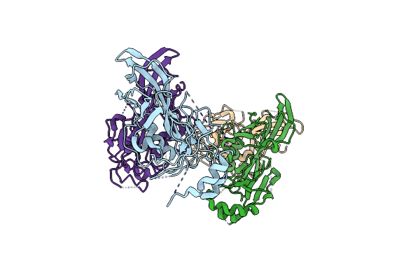
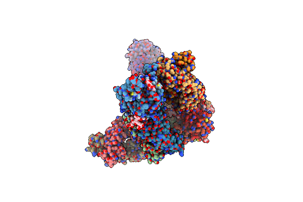
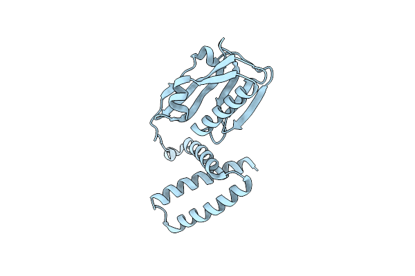
Organism: Homo sapiens, Crimean-congo hemorrhagic fever virus (strain nigeria/ibar10200/1970)
Method: X-RAY DIFFRACTION Resolution:2.10 Å Release Date: 2021-12-01 Classification: VIRAL PROTEIN |
 |
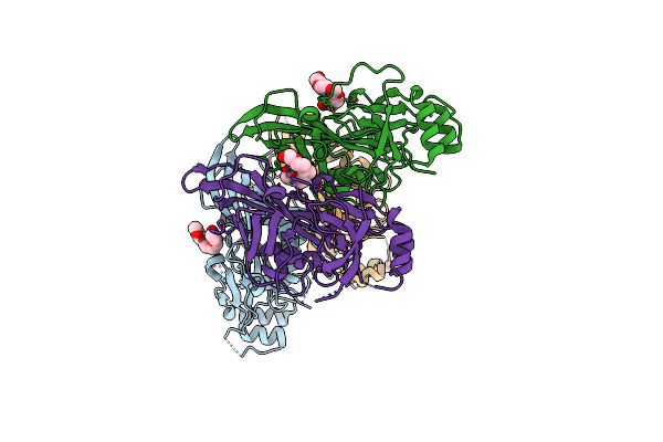
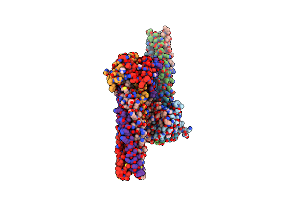
Crimean-Congo Hemorrhagic Fever Virus Envelope Glycoprotein Gc W1191H/W1197A/W1199A Mutant In Postfusion Conformation (Orthorhombic Crystal Form)
Organism: Crimean-congo hemorrhagic fever virus strain ibar10200
Method: X-RAY DIFFRACTION Resolution:2.20 Å Release Date: 2021-10-06 Classification: VIRAL PROTEIN Ligands: NAG, CL, PO4 |
 |
Crimean-Congo Hemorrhagic Fever Virus Envelope Glycoprotein Gc W1191H/W1197A/W1199A Mutant In Postfusion Conformation (Monoclinic Crystal Form)
Organism: Crimean-congo hemorrhagic fever virus (strain nigeria/ibar10200/1970)
Method: X-RAY DIFFRACTION Resolution:2.99 Å Release Date: 2021-10-06 Classification: VIRAL PROTEIN Ligands: NAG, CL |
 |
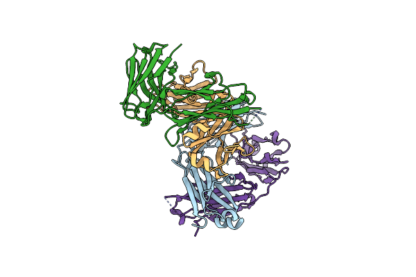
Structure Of The Niv F Glycoprotein In Complex With The 12B2 Neutralizing Antibody
Organism: Nipah virus, Mus musculus
Method: ELECTRON MICROSCOPY Release Date: 2021-05-05 Classification: VIRAL PROTEIN/IMMUNE SYSTEM |
 |
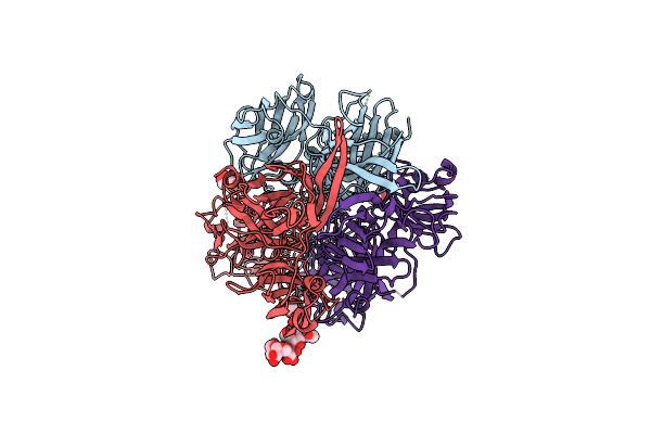
Structure Of The Hev F Glycoprotein In Complex With The 1F5 Neutralizing Antibody
Organism: Hendra henipavirus, Mus musculus
Method: ELECTRON MICROSCOPY Release Date: 2021-05-05 Classification: VIRAL PROTEIN/IMMUNE SYSTEM Ligands: NAG |
 |
Crystal Structure Demonstrating Ctd-Ctd Interactions Of Zaire Ebola Virus Vp40 Dimer
Organism: Zaire ebolavirus (strain mayinga-76)
Method: X-RAY DIFFRACTION Resolution:2.46 Å Release Date: 2020-10-28 Classification: VIRAL PROTEIN Ligands: 1PE |
 |
Low Resolution Crystal Structure Of Zaire Ebola Virus Vp40 In Space Group P6422
Organism: Zaire ebolavirus (strain mayinga-76)
Method: X-RAY DIFFRACTION Resolution:3.77 Å Release Date: 2020-10-21 Classification: VIRAL PROTEIN |
 |
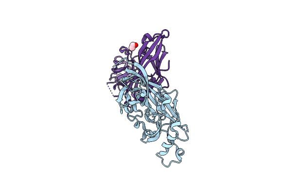
Organism: Crimean-congo hemorrhagic fever orthonairovirus
Method: X-RAY DIFFRACTION Resolution:2.52 Å Release Date: 2020-02-05 Classification: VIRAL PROTEIN Ligands: NAG |
 |
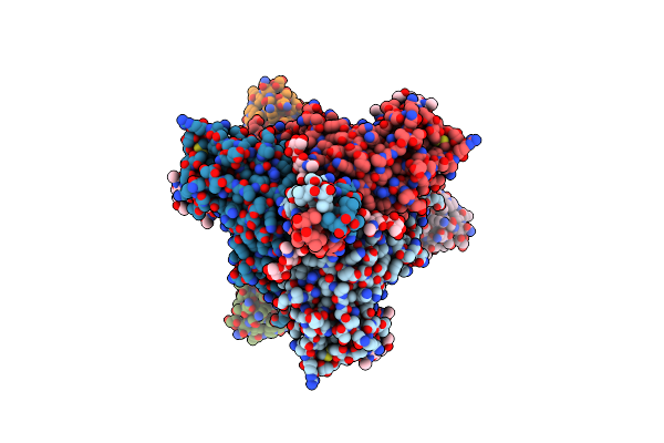
Ebola Virus Makona Variant Gp (Mucin-Deleted) In Complex With Pan-Ebolavirus Human Antibody Adi-15878 Fab
Organism: Zaire ebolavirus, Homo sapiens
Method: ELECTRON MICROSCOPY Release Date: 2018-09-12 Classification: VIRAL PROTEIN Ligands: NAG |
 |
Bundibugyo Virus Gp (Mucin-Deleted) In Complex With Pan-Ebolavirus Human Antibody Adi-15878 Fab
Organism: Bundibugyo ebolavirus, Homo sapiens
Method: ELECTRON MICROSCOPY Release Date: 2018-09-12 Classification: VIRAL PROTEIN Ligands: NAG |
 |
Organism: Homo sapiens
Method: X-RAY DIFFRACTION Resolution:2.10 Å Release Date: 2018-09-12 Classification: IMMUNE SYSTEM Ligands: CL, AE3, PG4 |
 |
Crystal Structure Of Marburg Virus Gp In Complex With The Human Survivor Antibody Mr78
Organism: Lake victoria marburgvirus (strain ravn-87), Homo sapiens
Method: X-RAY DIFFRACTION Resolution:3.60 Å Release Date: 2017-03-01 Classification: VIRAL PROTEIN/IMMUNE SYSTEM Ligands: NAG, MAN |
 |
Organism: Ebola virus sp., Zaire ebolavirus, Homo sapiens
Method: X-RAY DIFFRACTION Resolution:3.30 Å Release Date: 2016-03-09 Classification: VIRAL PROTEIN/IMMUNE SYSTEM |
 |
Organism: Lake victoria marburgvirus (strain musoke-80)
Method: X-RAY DIFFRACTION Resolution:2.81 Å Release Date: 2016-01-20 Classification: VIRAL PROTEIN Ligands: EOH |
 |
Organism: Marburg virus - musoke, kenya, 1980
Method: X-RAY DIFFRACTION Resolution:2.65 Å Release Date: 2014-03-26 Classification: VIRAL PROTEIN |
 |
Organism: Influenza a virus
Method: X-RAY DIFFRACTION Resolution:2.70 Å Release Date: 2014-02-19 Classification: VIRAL PROTEIN |
 |
Organism: Influenza a virus
Method: X-RAY DIFFRACTION Resolution:3.16 Å Release Date: 2014-02-19 Classification: VIRAL PROTEIN |
 |
Organism: Nipah virus
Method: X-RAY DIFFRACTION Resolution:2.20 Å Release Date: 2013-11-27 Classification: VIRAL PROTEIN Ligands: IMD |
 |
Organism: Sudan ebolavirus
Method: X-RAY DIFFRACTION Resolution:1.83 Å Release Date: 2013-08-21 Classification: VIRAL PROTEIN |