
Search Count: 32
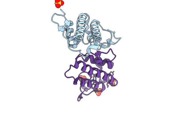 |
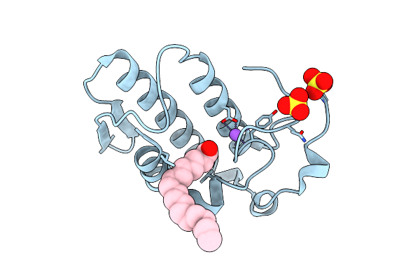
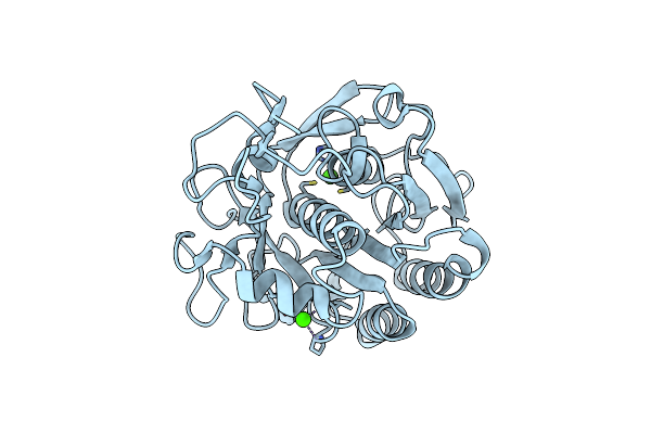
Crystal Structure Of Heterodimeric Crotoxin B From Crotalus Durissus Collilineatus
Organism: Crotalus durissus collilineatus
Method: X-RAY DIFFRACTION Resolution:1.89 Å Release Date: 2026-01-28 Classification: HYDROLASE Ligands: SO4 |
 |
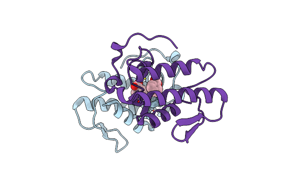
Organism: Arabidopsis thaliana
Method: X-RAY DIFFRACTION Resolution:3.51 Å Release Date: 2025-08-13 Classification: PROTEIN TRANSPORT Ligands: MAN, NAG |
 |
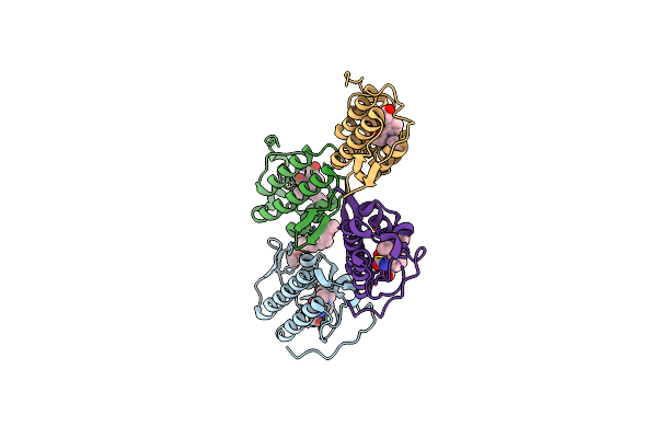
Organism: Bothrops jararacussu
Method: X-RAY DIFFRACTION Resolution:2.10 Å Release Date: 2022-11-09 Classification: TOXIN Ligands: 85O |
 |
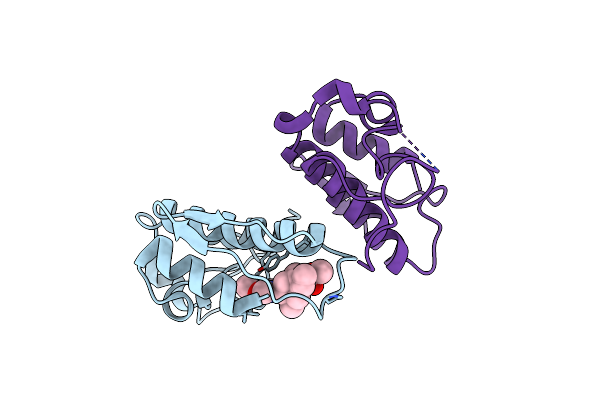
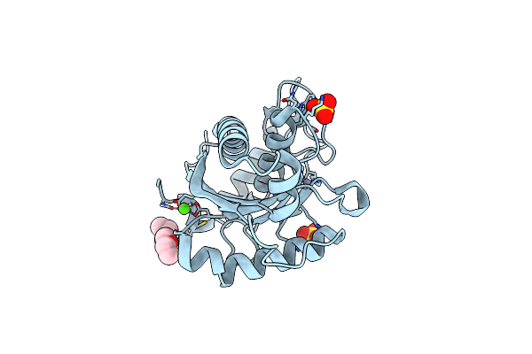
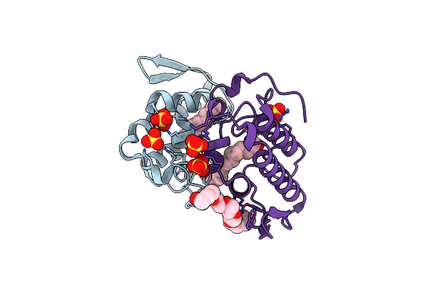
Crystal Structure Of The Am0627 (E326A) Inactive Mutant In Complex With Psgl-1-Like Bis-T Glycopeptide And Zn2+
Organism: Akkermansia muciniphila, Synthetic construct
Method: X-RAY DIFFRACTION Resolution:1.50 Å Release Date: 2022-07-20 Classification: HYDROLASE Ligands: GOL, ZN |
 |
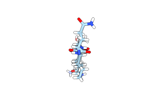
Microed Structure Of Human Zinc Finger Protein 292 Segment (534-542) Phased By Arcimboldo-Borges
Organism: Homo sapiens
Method: ELECTRON CRYSTALLOGRAPHY Resolution:1.50 Å Release Date: 2022-06-01 Classification: PROTEIN FIBRIL Ligands: DMS |
 |
Organism: Homo sapiens
Method: ELECTRON CRYSTALLOGRAPHY Resolution:1.00 Å Release Date: 2022-06-01 Classification: PROTEIN FIBRIL |
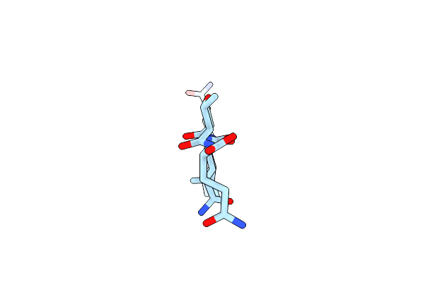 |
Microed Structure Of Human Cpeb3 Segment (154-161) Straight Polymorph Phased By Arcimboldo-Borges
Organism: Homo sapiens
Method: ELECTRON CRYSTALLOGRAPHY Resolution:1.20 Å Release Date: 2022-06-01 Classification: PROTEIN FIBRIL |
 |
Microed Structure Of Human Cpeb3 Segment(154-161) Kinked Polymorph Phased By Arcimboldo-Borges
Organism: Homo sapiens
Method: ELECTRON CRYSTALLOGRAPHY Resolution:1.20 Å Release Date: 2022-06-01 Classification: PROTEIN FIBRIL Ligands: HOH |
 |
X-Ray Structure Of A Sequence Variant Of A Repeat Segment Of The Yeast Prion New1P
Organism: Saccharomyces cerevisiae
Method: X-RAY DIFFRACTION Resolution:1.10 Å Release Date: 2022-06-01 Classification: PROTEIN FIBRIL |
 |
Organism: Homo sapiens
Method: ELECTRON CRYSTALLOGRAPHY Resolution:1.40 Å Release Date: 2022-06-01 Classification: PROTEIN FIBRIL |
 |
Microed Structure Of A Mutant Mammalian Prion Segment Phased By Arcimboldo-Borges
Organism: Homo sapiens
Method: ELECTRON CRYSTALLOGRAPHY Resolution:1.50 Å Release Date: 2022-06-01 Classification: PROTEIN FIBRIL Ligands: HOH |
 |
Microed Structure Of Sequence Variant Of Repeat Segment Of The Yeast Prion New1P Phased By Arcimboldo-Borges
Organism: Saccharomyces cerevisiae
Method: ELECTRON CRYSTALLOGRAPHY Resolution:1.30 Å Release Date: 2022-06-01 Classification: PROTEIN FIBRIL |
 |
Organism: Synthetic construct
Method: ELECTRON CRYSTALLOGRAPHY Resolution:0.90 Å Release Date: 2022-06-01 Classification: PROTEIN FIBRIL |
 |
Bthtx-Ii Variant B, From Bothrops Jararacussu Venom, Complexed With Stearic Acid
Organism: Bothrops jararacussu
Method: X-RAY DIFFRACTION Resolution:1.71 Å Release Date: 2022-01-05 Classification: TOXIN Ligands: STE, NA, SO4 |
 |
Bthtx-Ii Variant A, From Bothrops Jararacussu Venom, Complexed With Benzoic Acid
Organism: Bothrops jararacussu
Method: X-RAY DIFFRACTION Resolution:1.70 Å Release Date: 2022-01-05 Classification: TOXIN Ligands: BEZ |
 |
Myotoxin I From Bothrops Moojeni Co-Crystallized With Synthetic Inhibitor Varespladib (Ly315920)
Organism: Bothrops moojeni
Method: X-RAY DIFFRACTION Resolution:1.76 Å Release Date: 2021-05-19 Classification: TOXIN Ligands: VRD, PE4 |
 |
Crystal Structure O Bmoomp-I, A P-I Metalloproteinase From Bothrops Moojeni
Organism: Bothrops moojeni
Method: X-RAY DIFFRACTION Resolution:1.92 Å Release Date: 2020-10-07 Classification: HYDROLASE Ligands: CA, ZN, PG4, SO4 |
 |
Crystal Structure Of C. Parvum Gna1 In Complex With Acetyl-Coa And Glucose 6P.
Organism: Cryptosporidium parvum iowa ii
Method: X-RAY DIFFRACTION Resolution:1.95 Å Release Date: 2020-09-30 Classification: TRANSFERASE Ligands: ACO, G6P |
 |
Organism: Parengyodontium album
Method: ELECTRON CRYSTALLOGRAPHY Resolution:1.60 Å Release Date: 2020-08-12 Classification: HYDROLASE Ligands: CA |
 |
Crystal Structure Of Myotoxin Ii From Bothrops Moojeni Complexed To Myristic Acid
Organism: Bothrops moojeni
Method: X-RAY DIFFRACTION Resolution:1.95 Å Release Date: 2018-10-03 Classification: TOXIN Ligands: MYR, SO4, PE4 |