
Search Count: 28
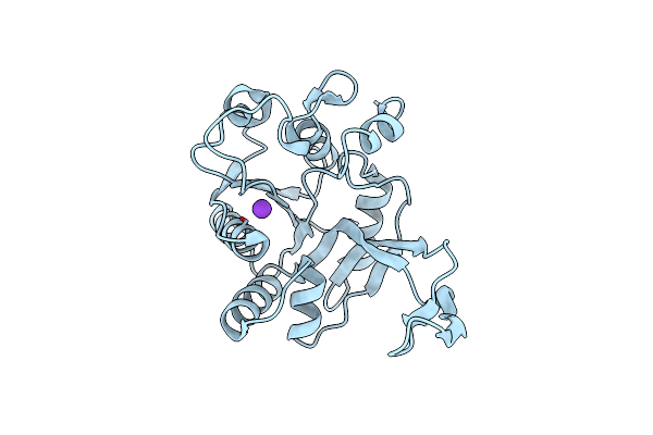 |
Organism: Pseudomonas aeruginosa
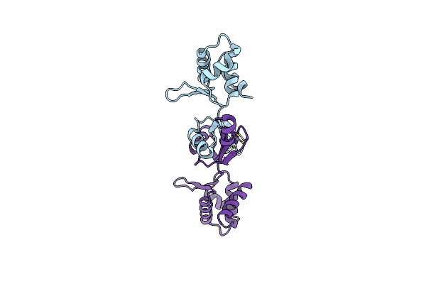
Method: X-RAY DIFFRACTION Release Date: 2025-06-11 Classification: TRANSFERASE Ligands: K |
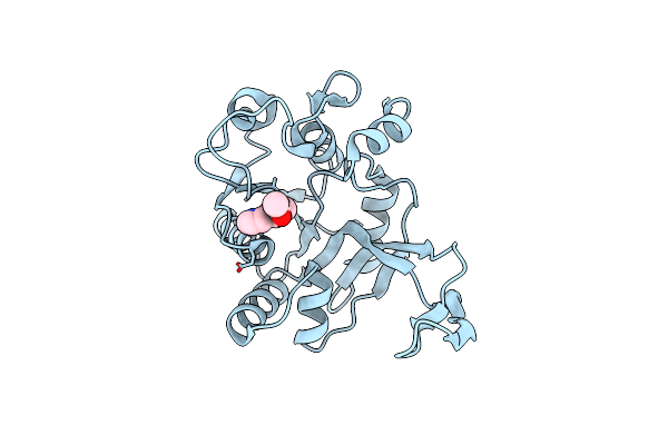 |
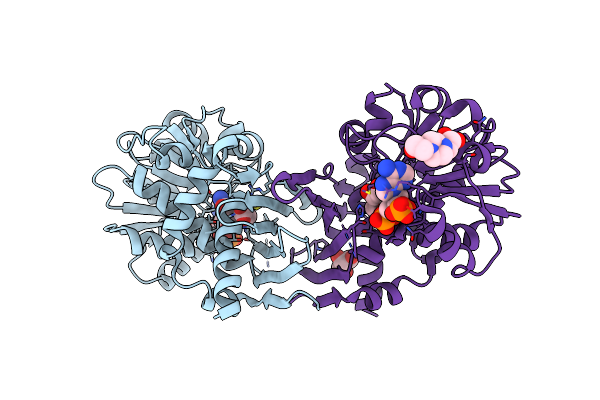
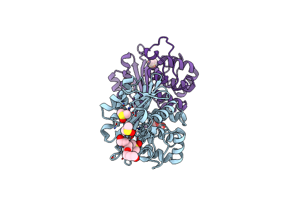
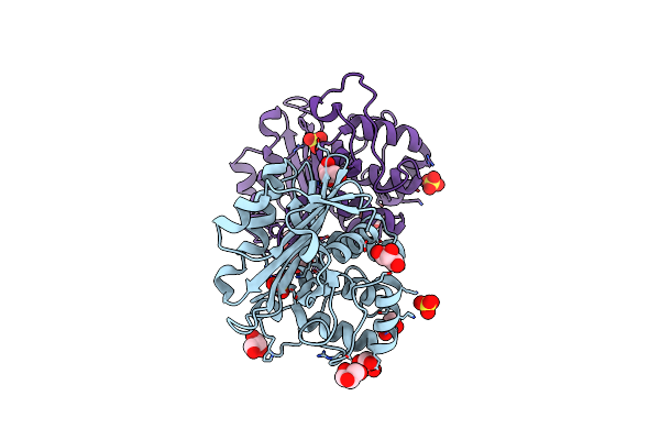
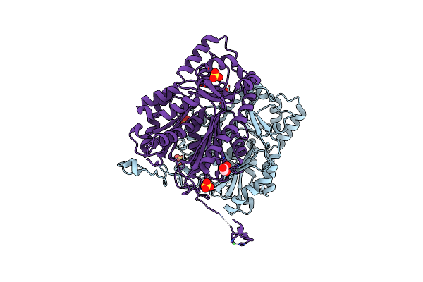
Crystal Structure Of Pseudomonas Aeruginosa Ispd In Complex With C11H12N2O3
Organism: Pseudomonas aeruginosa
Method: X-RAY DIFFRACTION Release Date: 2025-06-11 Classification: TRANSFERASE Ligands: A1IJS |
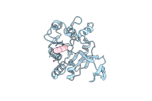 |
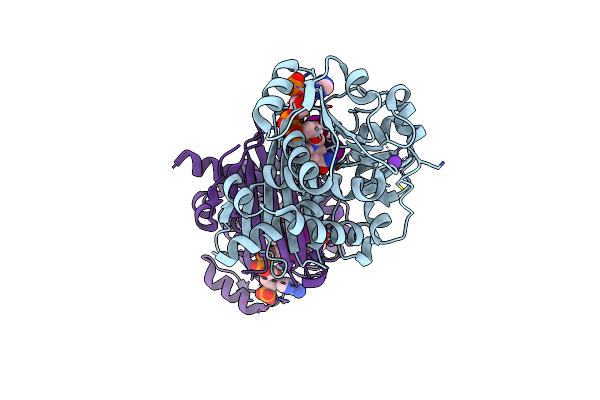
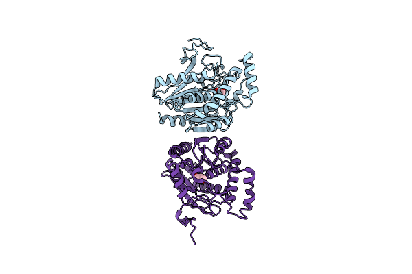
Organism: Pseudomonas aeruginosa
Method: X-RAY DIFFRACTION Release Date: 2025-06-11 Classification: TRANSFERASE Ligands: A1IJT |
 |
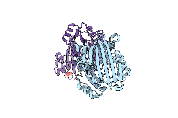
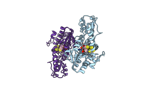
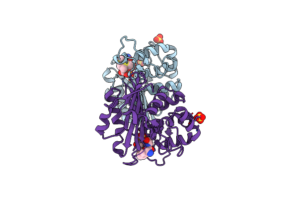
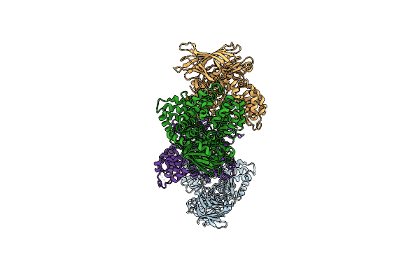
E.Coli Seryl-Trna Synthetase (Arm Deletion Mutant) Bound To Sulphamoyl Seryl-Adenylate Analogue
Organism: Escherichia coli k-12
Method: X-RAY DIFFRACTION Resolution:2.32 Å Release Date: 2025-05-14 Classification: TRANSLATION Ligands: MG, SO4, SSA |
 |
Organism: Mus musculus
Method: X-RAY DIFFRACTION Resolution:2.30 Å Release Date: 2014-02-05 Classification: OXIDOREDUCTASE Ligands: K, GOL |
 |
Organism: Mus musculus
Method: X-RAY DIFFRACTION Resolution:2.19 Å Release Date: 2014-02-05 Classification: OXIDOREDUCTASE Ligands: NDP, PYR, EPE, GOL |
 |
Organism: Mus musculus
Method: X-RAY DIFFRACTION Resolution:1.75 Å Release Date: 2014-02-05 Classification: OXIDOREDUCTASE Ligands: NDP, T3, K |
 |
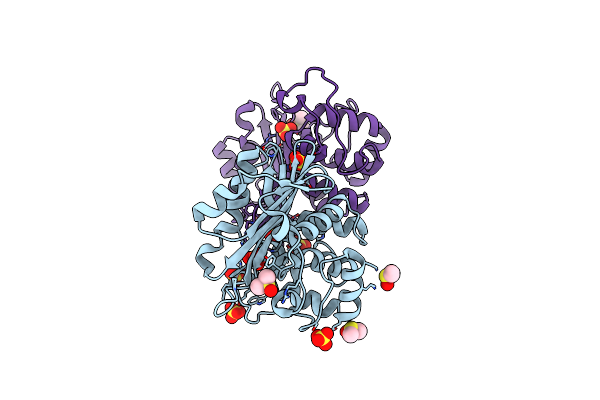
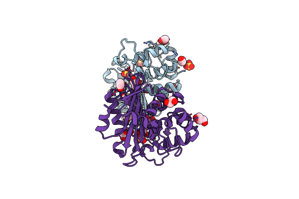
Crystal Structures Of Escherichia Coli Isph In Complex With Ambpp A Potent Inhibitor Of The Methylerythritol Phosphate Pathway
Organism: Escherichia coli
Method: X-RAY DIFFRACTION Resolution:1.68 Å Release Date: 2013-01-09 Classification: OXIDOREDUCTASE Ligands: SF4, 10E |
 |
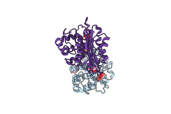
Crystal Structures Of Escherichia Coli Isph In Complex With Tmbpp A Potent Inhibitor Of The Methylerythritol Phosphate Pathway
Organism: Escherichia coli
Method: X-RAY DIFFRACTION Resolution:1.95 Å Release Date: 2013-01-09 Classification: OXIDOREDUCTASE Ligands: 10G, SF4 |
 |
Organism: Pseudomonas aeruginosa
Method: X-RAY DIFFRACTION Resolution:1.90 Å Release Date: 2010-12-08 Classification: HYDROLASE Ligands: SO4, DMS, PG4 |
 |
Crystal Structure Of The Class D Beta-Lactamase Oxa-10 At 1.35 A Resolution
Organism: Pseudomonas aeruginosa
Method: X-RAY DIFFRACTION Resolution:1.35 Å Release Date: 2010-12-08 Classification: HYDROLASE Ligands: SO4, PG4, DMS, GOL |
 |
Organism: Pseudomonas aeruginosa
Method: X-RAY DIFFRACTION Resolution:1.79 Å Release Date: 2010-08-25 Classification: HYDROLASE Ligands: ZZ7, SO4 |
 |
Organism: Pseudomonas aeruginosa
Method: X-RAY DIFFRACTION Resolution:2.10 Å Release Date: 2010-08-25 Classification: HYDROLASE Ligands: SO4, GOL, EDO |
 |
Crystal Structure Of The Oxa-10 V117T Mutant At Ph 6.5 Inhibited By A Chloride Ion
Organism: Pseudomonas aeruginosa
Method: X-RAY DIFFRACTION Resolution:1.80 Å Release Date: 2010-05-19 Classification: HYDROLASE Ligands: GOL, CL, CIT, SO4 |
 |
Organism: Pseudomonas aeruginosa
Method: X-RAY DIFFRACTION Resolution:1.80 Å Release Date: 2010-05-19 Classification: HYDROLASE Ligands: SO4, GOL |
 |
Organism: Homo sapiens
Method: X-RAY DIFFRACTION Resolution:2.20 Å Release Date: 2009-12-08 Classification: HYDROLASE Ligands: GOL |
 |
Organism: Bacillus subtilis
Method: X-RAY DIFFRACTION Resolution:2.05 Å Release Date: 2008-10-14 Classification: DNA BINDING PROTEIN Ligands: ZN |
 |
Organism: Colwellia psychrerythraea
Method: X-RAY DIFFRACTION Resolution:2.70 Å Release Date: 2008-07-01 Classification: HYDROLASE Ligands: ZN |
 |
Crystal Structure Of The Ligand Binding Domain Of Polyandrocarpa Misakiensis Rxr In Tetramer In Absence Of Ligand
Organism: Polyandrocarpa misakiensis
Method: X-RAY DIFFRACTION Resolution:2.90 Å Release Date: 2008-05-27 Classification: TRANSCRIPTION |
 |
Crystal Structure Of Marchantia Polymorpha Stilbenecarboxylate Synthase 2 (Stcs2)
Organism: Marchantia polymorpha
Method: X-RAY DIFFRACTION Resolution:1.90 Å Release Date: 2007-03-13 Classification: TRANSFERASE Ligands: SO4, GOL, NI |