
Search Count: 20
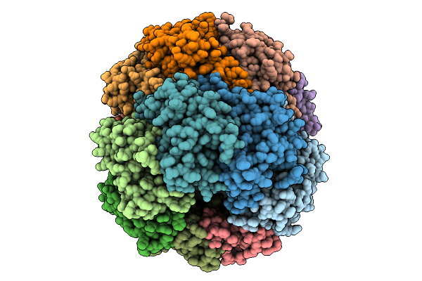 |
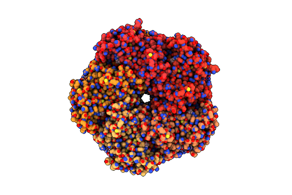
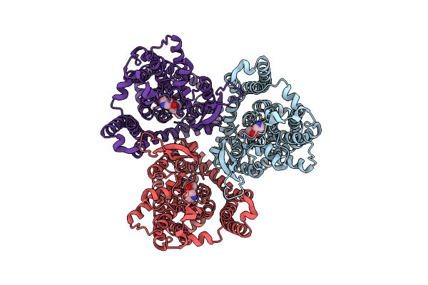
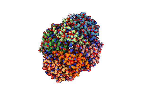
Crystal Structure Of Neisseria Meningitidis Clpp Protease Complex With Boronate Compound Bc8A
Organism: Neisseria meningitidis
Method: X-RAY DIFFRACTION Release Date: 2025-09-03 Classification: HYDROLASE Ligands: JT7 |
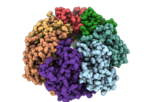 |
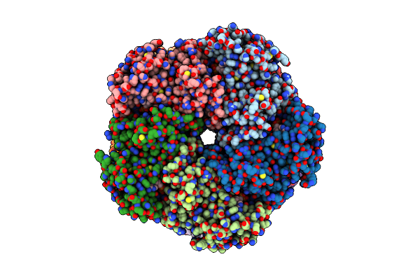
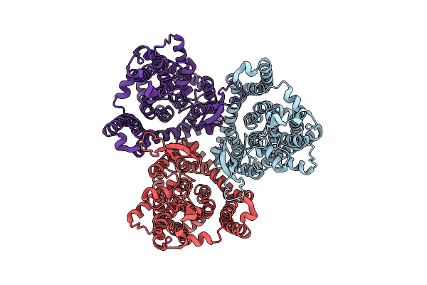
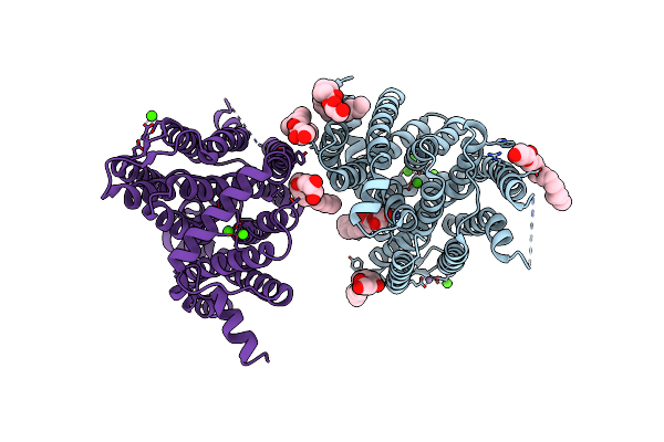
Crystal Structure Of Neisseria Meningitidis Clpp Protease Complex With Noncovalent Activator, Acp1-01
Organism: Neisseria meningitidis
Method: X-RAY DIFFRACTION Release Date: 2025-09-03 Classification: HYDROLASE Ligands: A1BLZ |
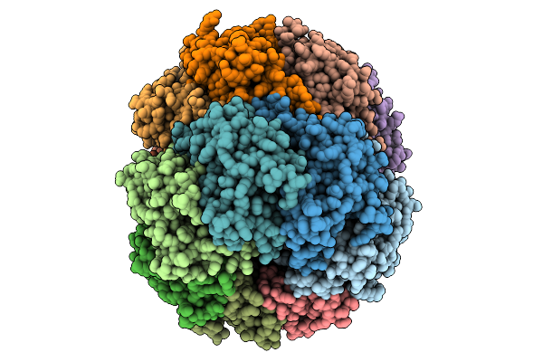 |
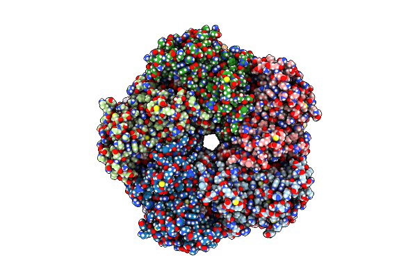
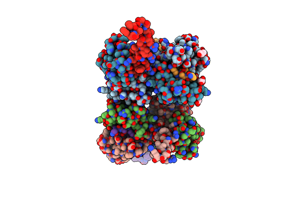
Crystal Structure Of Neisseria Meningitidis Clpp Protease Complex With Small Molecule Activator, Dioctatin
Organism: Neisseria meningitidis
Method: X-RAY DIFFRACTION Release Date: 2025-09-03 Classification: HYDROLASE Ligands: A1BMH |
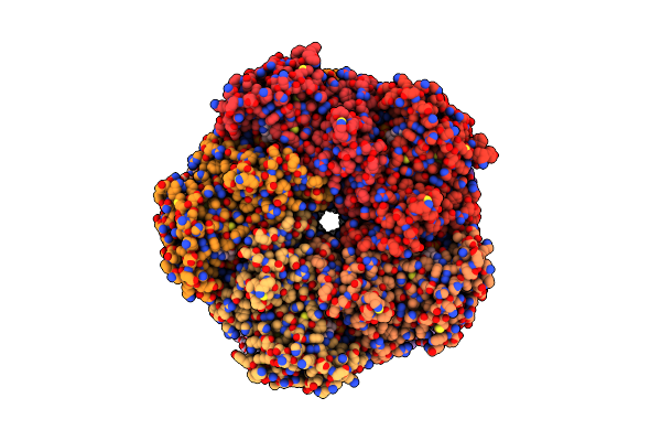 |
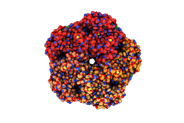
Organism: Homo sapiens
Method: ELECTRON MICROSCOPY Release Date: 2025-07-02 Classification: LIGASE Ligands: ATP, MG, GLN |
 |
Organism: Homo sapiens
Method: ELECTRON MICROSCOPY Release Date: 2025-07-02 Classification: LIGASE Ligands: ATP, MG |
 |
Organism: Homo sapiens
Method: ELECTRON MICROSCOPY Release Date: 2025-07-02 Classification: LIGASE Ligands: ADP, MG |
 |
Organism: Homo sapiens
Method: ELECTRON MICROSCOPY Release Date: 2025-07-02 Classification: LIGASE Ligands: ADP, MG |
 |
Organism: Homo sapiens
Method: ELECTRON MICROSCOPY Release Date: 2025-07-02 Classification: LIGASE Ligands: MG |
 |
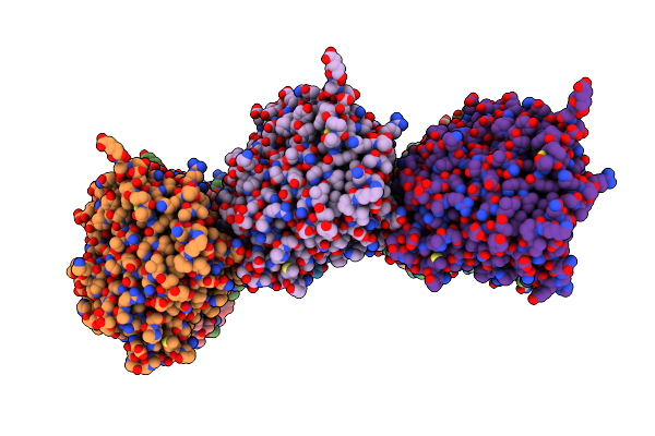
Crystal Structure Of Human Ace2 Bound To The Spike Receptor-Binding Domain From A Cave Bat Sarbecovirus Closely Related To Sars-Cov-2.
Organism: Homo sapiens, Sarbecovirus
Method: X-RAY DIFFRACTION Resolution:2.94 Å Release Date: 2022-01-19 Classification: VIRAL PROTEIN Ligands: NAG, SO4, TRS, PEG, ZN, CL, GOL |
 |
Organism: Serendipita indica
Method: X-RAY DIFFRACTION Resolution:2.90 Å Release Date: 2021-11-17 Classification: TRANSPORT PROTEIN Ligands: PO4, BNG |
 |
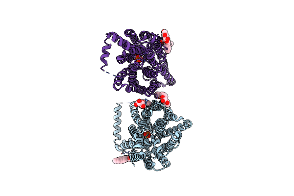
Asct2 In The Presence Of The Inhibitor Lc-Bpe (Position "Up") In The Outward-Open Conformation.
Organism: Homo sapiens
Method: ELECTRON MICROSCOPY Release Date: 2021-09-22 Classification: MEMBRANE PROTEIN Ligands: TG2 |
 |
Asct2 In The Presence Of The Inhibitor Lc-Bpe (Position "Down") In The Outward-Open Conformation.
Organism: Homo sapiens
Method: ELECTRON MICROSCOPY Release Date: 2021-09-22 Classification: MEMBRANE PROTEIN Ligands: TJ5 |
 |
Asct2 In The Presence Of The Inhibitor Era-21 In The Outward-Open Conformation.
Organism: Homo sapiens
Method: ELECTRON MICROSCOPY Release Date: 2021-09-22 Classification: MEMBRANE PROTEIN |
 |
Organism: Sus scrofa
Method: ELECTRON MICROSCOPY Release Date: 2019-05-22 Classification: STRUCTURAL PROTEIN Ligands: GTP, MG, GDP |
 |
Organism: Sus scrofa
Method: ELECTRON MICROSCOPY Release Date: 2019-05-22 Classification: STRUCTURAL PROTEIN Ligands: GTP, MG, GDP |
 |
Organism: Sus scrofa
Method: ELECTRON MICROSCOPY Release Date: 2019-05-22 Classification: STRUCTURAL PROTEIN Ligands: GTP, MG, GDP |
 |
Organism: Sus scrofa
Method: ELECTRON MICROSCOPY Release Date: 2019-05-22 Classification: STRUCTURAL PROTEIN Ligands: GTP, MG, GDP |
 |
Organism: Staphylococcus aureus subsp. aureus str. newman
Method: ELECTRON MICROSCOPY Release Date: 2018-06-27 Classification: HYDROLASE |
 |
Organism: Saccharomyces cerevisiae
Method: X-RAY DIFFRACTION Resolution:2.30 Å Release Date: 2013-05-08 Classification: MEMBRANE PROTEIN/METAL TRANSPORT Ligands: OLC, CA, MN, IOD |
 |
Crystal Structure Of Yeast Calmodulin Bound To The C-Terminal Fragment Of Spindle Pole Body Protein Spc110
Organism: Kluyveromyces lactis, Saccharomyces cerevisiae s288c
Method: X-RAY DIFFRACTION Resolution:2.15 Å Release Date: 2012-03-07 Classification: PROTEIN BINDING Ligands: SR, GOL, SO4 |