
Search Count: 38
 |
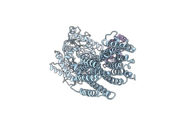
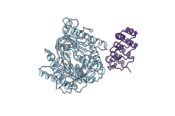
Kalium Channelrhodopsin 1 C110A Mutant From Hyphochytrium Catenoides, Dark State
Organism: Hyphochytrium catenoides
Method: ELECTRON MICROSCOPY Release Date: 2025-02-19 Classification: MEMBRANE PROTEIN Ligands: RET, CLR, PEE |
 |
Kalium Channelrhodopsin 1 C110A Mutant From Hyphochytrium Catenoides, Laser-Flash-Illuminated
Organism: Hyphochytrium catenoides
Method: ELECTRON MICROSCOPY Release Date: 2025-02-19 Classification: MEMBRANE PROTEIN Ligands: RET, CLR, PEE |
 |
Kalium Channelrhodopsin 1 C110A Mutant From Hyphochytrium Catenoides, Continuous Illumination State
Organism: Hyphochytrium catenoides
Method: ELECTRON MICROSCOPY Release Date: 2025-02-19 Classification: MEMBRANE PROTEIN Ligands: RET, CLR, PEE |
 |
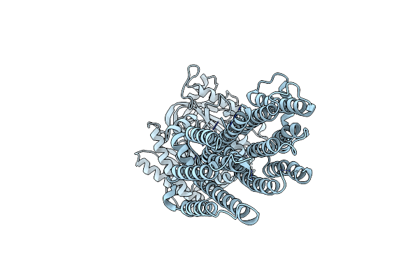
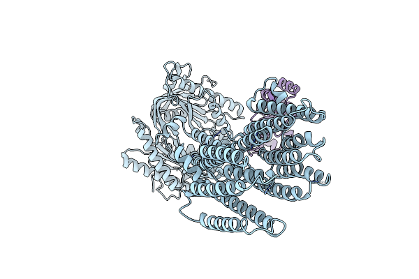
Kalium Channelrhodopsin 1 From Hyphochytrium Catenoides (Hckcr1) Embedded In Peptidisc
Organism: Hyphochytrium catenoides
Method: ELECTRON MICROSCOPY Release Date: 2023-07-26 Classification: TRANSPORT PROTEIN Ligands: RET, NA, CLR, PEE |
 |
Cation Channelrhodopsin From Hyphochytrium Catenoides (Hcccr) Embedded In Peptidisc
Organism: Hyphochytrium catenoides
Method: ELECTRON MICROSCOPY Release Date: 2023-07-26 Classification: TRANSPORT PROTEIN Ligands: RET, NA, CLR, PEE |
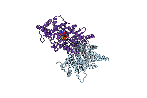 |
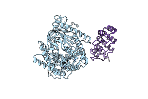
Focus Refinement Of Soluble Domain Of Adenylyl Cyclase 9 In Complex With Gs Protein Alpha Subunit And Mant-Gtp
Organism: Bos taurus
Method: ELECTRON MICROSCOPY Release Date: 2022-01-26 Classification: SIGNALING PROTEIN Ligands: GSP, MG |
 |
Organism: Bos taurus
Method: ELECTRON MICROSCOPY Release Date: 2022-01-19 Classification: SIGNALING PROTEIN |
 |
Organism: Bos taurus, Synthetic construct
Method: ELECTRON MICROSCOPY Release Date: 2022-01-19 Classification: SIGNALING PROTEIN |
 |
Focus Refinement Of Soluble Domain Of Adenylyl Cyclase 9 In Complex With Darpin C4 And Mant-Gtp
Organism: Bos taurus, Synthetic construct
Method: ELECTRON MICROSCOPY Release Date: 2022-01-19 Classification: SIGNALING PROTEIN |
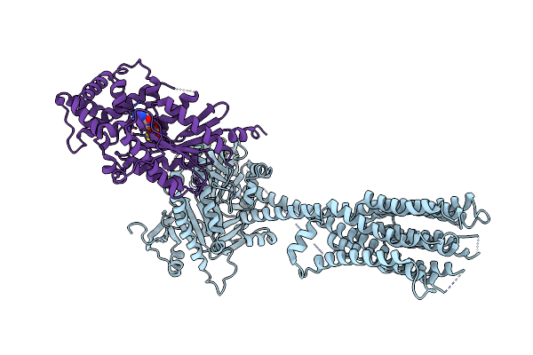 |
Structure Of Adenylyl Cyclase 9 In Complex With Gs Protein Alpha Subunit And Mant-Gtp
Organism: Bos taurus
Method: ELECTRON MICROSCOPY Release Date: 2022-01-19 Classification: SIGNALING PROTEIN Ligands: GSP, MG |
 |
Organism: Bos taurus, Synthetic construct
Method: ELECTRON MICROSCOPY Release Date: 2022-01-19 Classification: SIGNALING PROTEIN |
 |
Organism: Bos taurus, Synthetic construct
Method: ELECTRON MICROSCOPY Release Date: 2022-01-19 Classification: SIGNALING PROTEIN |
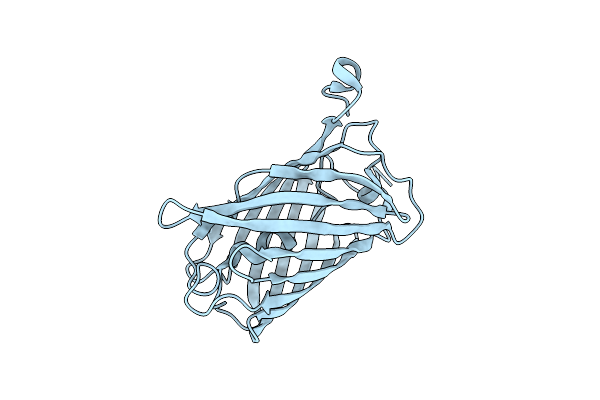 |
Organism: Discosoma sp.
Method: X-RAY DIFFRACTION Resolution:1.60 Å Release Date: 2020-12-16 Classification: FLUORESCENT PROTEIN Ligands: CL |
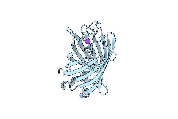 |
Mturquoise2 Sg P212121 - Directional Optical Properties Of Fluorescent Proteins
Organism: Vaccinia virus
Method: X-RAY DIFFRACTION Resolution:1.85 Å Release Date: 2020-12-16 Classification: FLUORESCENT PROTEIN Ligands: K |
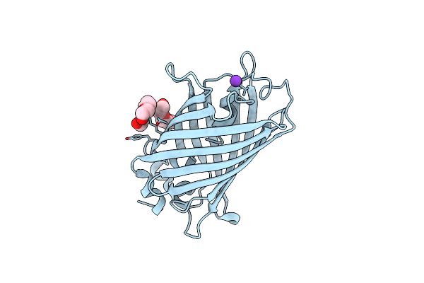 |
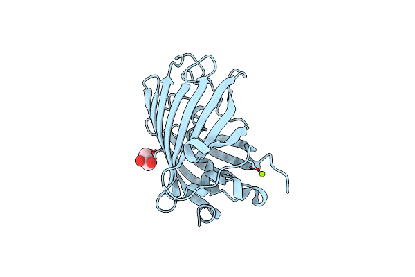
Organism: Synthetic construct
Method: X-RAY DIFFRACTION Resolution:1.70 Å Release Date: 2020-12-16 Classification: FLUORESCENT PROTEIN Ligands: K, PG4 |
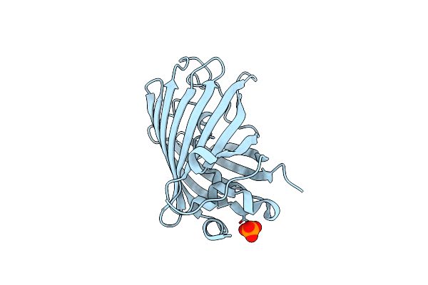 |
Egfp_In_Acidic_Env Directionality Of Optical Properties Of Fluorescent Proteins
Organism: Synthetic construct
Method: X-RAY DIFFRACTION Resolution:1.55 Å Release Date: 2020-12-16 Classification: FLUORESCENT PROTEIN Ligands: PO4 |
 |
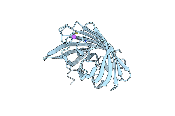
Egfp In Neutral Ph, Directionality Of Optical Properties Of Fluorescent Proteins
Organism: Synthetic construct
Method: X-RAY DIFFRACTION Resolution:1.65 Å Release Date: 2020-12-16 Classification: FLUORESCENT PROTEIN Ligands: GOL, MG |
 |
Organism: Lobophyllia hemprichii
Method: X-RAY DIFFRACTION Resolution:1.55 Å Release Date: 2020-12-16 Classification: FLUORESCENT PROTEIN Ligands: CL, NA |
 |
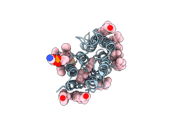
Crystal Structure Of The Selenomethionine-Substituted Iron-Regulated Protein Frpd From Neisseria Meningitidis
Organism: Neisseria meningitidis
Method: X-RAY DIFFRACTION Resolution:1.40 Å Release Date: 2017-02-01 Classification: UNKNOWN FUNCTION Ligands: P6G, 1PE, NA, AZI |
 |
Crystal Structure Of The Neisseria Meningitidis Iron-Regulated Outer Membrane Lipoprotein Frpd
Organism: Neisseria meningitidis mc58
Method: X-RAY DIFFRACTION Resolution:2.30 Å Release Date: 2017-02-01 Classification: UNKNOWN FUNCTION |