
Search Count: 26
 |
Organism: Streptomyces cacaoi
Method: X-RAY DIFFRACTION Release Date: 2025-10-22 Classification: OXIDOREDUCTASE Ligands: ZN, GOL |
 |
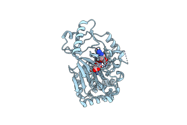
X-Ray Crystal Structure Of Clavaminate Synthase With Vanadyl, Succinate, And Deoxyproclavaminic Acid
Organism: Streptomyces antibioticus
Method: X-RAY DIFFRACTION Resolution:1.50 Å Release Date: 2021-02-24 Classification: OXIDOREDUCTASE Ligands: VVO, SIN, RQJ |
 |
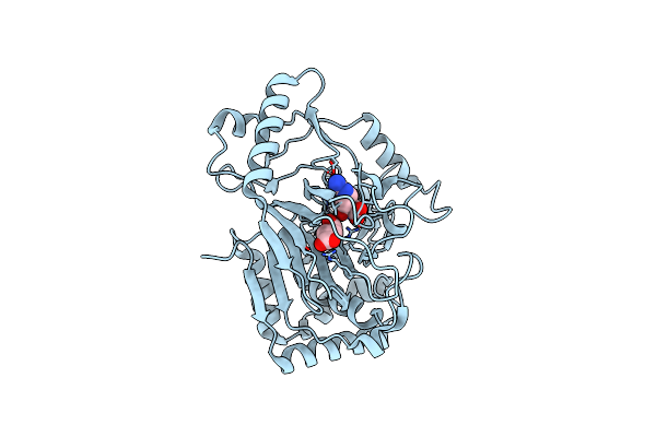
X-Ray Structure Of Clavaminate Synthase With Vanadyl, Succinate, And Deoxyguanidinoproclavaminic Acid
Organism: Streptomyces antibioticus
Method: X-RAY DIFFRACTION Resolution:1.50 Å Release Date: 2021-02-24 Classification: OXIDOREDUCTASE Ligands: SIN, PCX, VVO |
 |
X-Ray Crystal Structure Of Flavobacterium Johnsoniae Dimanganese(Ii) Ribonucleotide Reductase Beta Subunit (Aerobic)
Organism: Flavobacterium johnsoniae (strain atcc 17061 / dsm 2064 / uw101)
Method: X-RAY DIFFRACTION Resolution:1.87 Å Release Date: 2018-06-27 Classification: OXIDOREDUCTASE Ligands: MN, MG |
 |
X-Ray Crystal Structure Of Napi L-Arginine Desaturase Bound To Fe(Ii), L-Arginine, And Acetate
Organism: Streptomyces lusitanus
Method: X-RAY DIFFRACTION Resolution:2.10 Å Release Date: 2018-05-16 Classification: OXIDOREDUCTASE Ligands: FE2, ARG, ACT |
 |
X-Ray Crystal Structure Of Vioc Bound To Fe(Ii), L-Homoarginine, And 2-Oxoglutarate
Organism: Streptomyces vinaceus
Method: X-RAY DIFFRACTION Resolution:1.70 Å Release Date: 2018-05-16 Classification: OXIDOREDUCTASE Ligands: AKG, HRG, FE2 |
 |
X-Ray Crystal Structure Of Vioc Bound To Fe(Ii), 3S-Hydroxy-L-Homoarginine, And Succinate
Organism: Streptomyces vinaceus
Method: X-RAY DIFFRACTION Resolution:1.94 Å Release Date: 2018-05-16 Classification: OXIDOREDUCTASE Ligands: FE2, SIN, G3M |
 |
X-Ray Crystal Structure Of Vioc Bound To Vanadyl Ion, L-Homoarginine, And Succinate
Organism: Streptomyces vinaceus
Method: X-RAY DIFFRACTION Resolution:1.70 Å Release Date: 2018-05-16 Classification: OXIDOREDUCTASE Ligands: SIN, HRG, VVO, EDO |
 |
X-Ray Crystal Structure Of Flavobacterium Johnsoniae Dimanganese(Ii) Ribonucleotide Reductase Beta Subunit (Anaerobic)
Organism: Flavobacterium johnsoniae (strain atcc 17061 / dsm 2064 / uw101), Flavobacterium johnsoniae
Method: X-RAY DIFFRACTION Resolution:1.92 Å Release Date: 2018-04-18 Classification: OXIDOREDUCTASE Ligands: MN |
 |
X-Ray Crystal Structure Of Flavobacterium Johnsoniae Dimanganese(Ii) Ribonucleotide Reductase Beta Subunit (As-Isolated)
Organism: Flavobacterium johnsoniae (strain atcc 17061 / dsm 2064 / uw101)
Method: X-RAY DIFFRACTION Resolution:1.90 Å Release Date: 2018-04-18 Classification: OXIDOREDUCTASE Ligands: MN, MG |
 |
Vioc L-Arginine Hydroxylase Bound To Fe(Ii), L-Arginine, And 2-Oxo-Glutaric Acid
Organism: Streptomyces vinaceus
Method: X-RAY DIFFRACTION Resolution:1.60 Å Release Date: 2017-09-06 Classification: OXIDOREDUCTASE Ligands: FE2, AKG, ARG |
 |
Vioc L-Arginine Hydroxylase Bound To Fe(Ii), 3S-Hydroxy-L-Arginine, And 2Og
Organism: Streptomyces vinaceus
Method: X-RAY DIFFRACTION Resolution:1.80 Å Release Date: 2017-09-06 Classification: OXIDOREDUCTASE Ligands: AKG, FE2, ZZU |
 |
Vioc L-Arginine Hydroxylase Bound To Fe(Ii), L-Arginine, And A Peroxysuccinate Intermediate
Organism: Streptomyces vinaceus
Method: X-RAY DIFFRACTION Resolution:1.79 Å Release Date: 2017-09-06 Classification: OXIDOREDUCTASE Ligands: FE2, ARG, OKG |
 |
Vioc L-Arginine Hydroxylase Bound To Fe(Ii), 3S-Hydroxy-L-Arginine, And Succinate
Organism: Streptomyces vinaceus
Method: X-RAY DIFFRACTION Resolution:1.99 Å Release Date: 2017-09-06 Classification: OXIDOREDUCTASE Ligands: FE2, SIN, ZZU |
 |
Organism: Streptomyces vinaceus
Method: X-RAY DIFFRACTION Resolution:1.67 Å Release Date: 2017-09-06 Classification: OXIDOREDUCTASE Ligands: FE2, SIN, ARG |
 |
Vioc L-Arginine Hydroxylase Bound To The Vanadyl Ion, L-Arginine, And Succinate
Organism: Streptomyces vinaceus
Method: X-RAY DIFFRACTION Resolution:1.55 Å Release Date: 2017-09-06 Classification: OXIDOREDUCTASE Ligands: SIN, ARG, VVO |
 |
Structure Of Pas-Gaf Fragment Of Deinococcus Phytochrome By Serial Femtosecond Crystallography
Organism: Deinococcus radiodurans (strain atcc 13939 / dsm 20539 / jcm 16871 / lmg 4051 / nbrc 15346 / ncimb 9279 / r1 / vkm b-1422)
Method: X-RAY DIFFRACTION Resolution:1.65 Å Release Date: 2017-02-22 Classification: TRANSFERASE Ligands: LBV, NI, CL, EDO |
 |
Structure Of The Photosensory Module Of Deinococcus Phytochrome By Serial Femtosecond X-Ray Crystallography
Organism: Deinococcus radiodurans (strain atcc 13939 / dsm 20539 / jcm 16871 / lmg 4051 / nbrc 15346 / ncimb 9279 / r1 / vkm b-1422)
Method: X-RAY DIFFRACTION Resolution:3.30 Å Release Date: 2017-02-22 Classification: TRANSFERASE Ligands: LBV |
 |
Crystal Structure Of Carbapenem Synthase In Complex With (3S,5S)-Carbapenam
Organism: Pectobacterium carotovorum subsp. carotovorum
Method: X-RAY DIFFRACTION Resolution:2.10 Å Release Date: 2014-04-02 Classification: OXIDOREDUCTASE Ligands: FE2, AKG, GOL, 2TQ |
 |
Organism: Uncultured bacterium hf130_aepn_1
Method: X-RAY DIFFRACTION Resolution:1.85 Å Release Date: 2013-11-27 Classification: OXIDOREDUCTASE Ligands: FE, FLC |