
Search Count: 47
 |
Organism: Homo sapiens
Method: ELECTRON MICROSCOPY Release Date: 2025-09-03 Classification: CELL CYCLE Ligands: ZN |
 |
Organism: Homo sapiens
Method: ELECTRON MICROSCOPY Release Date: 2025-09-03 Classification: CELL CYCLE Ligands: ZN |
 |
Organism: Homo sapiens
Method: ELECTRON MICROSCOPY Release Date: 2025-09-03 Classification: CELL CYCLE Ligands: ZN |
 |
Organism: Homo sapiens
Method: ELECTRON MICROSCOPY Release Date: 2025-09-03 Classification: CELL CYCLE |
 |
Organism: Homo sapiens
Method: ELECTRON MICROSCOPY Release Date: 2025-09-03 Classification: CELL CYCLE Ligands: ZN |
 |
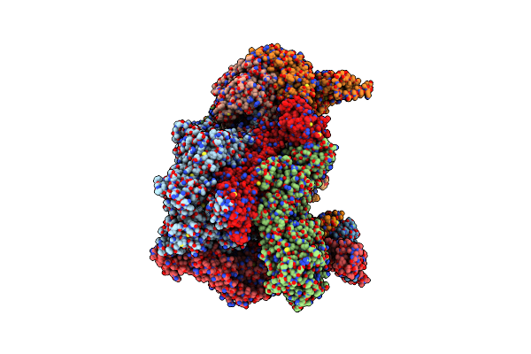
Cryo-Em Structure Of Human Separase Bound To Phosphorylated Scc1 (310-550 Aa)
Organism: Homo sapiens
Method: ELECTRON MICROSCOPY Release Date: 2025-09-03 Classification: CELL CYCLE Ligands: ZN |
 |
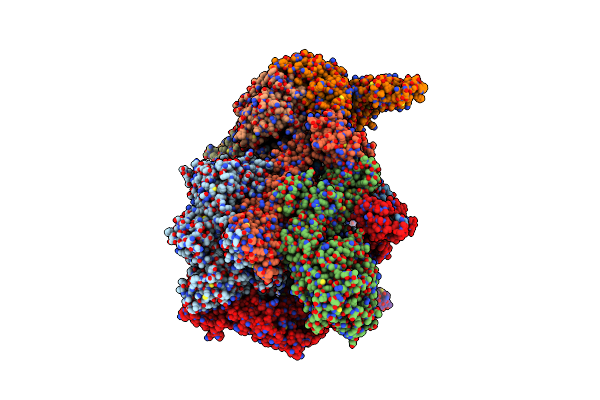
Cryo-Em Structure Of Human Separase Bound To Phosphorylated Scc1 (100-320 Aa)
Organism: Homo sapiens
Method: ELECTRON MICROSCOPY Release Date: 2025-09-03 Classification: CELL CYCLE Ligands: ZN |
 |
Organism: Lama glama
Method: X-RAY DIFFRACTION Resolution:2.85 Å Release Date: 2024-09-11 Classification: IMMUNE SYSTEM |
 |
High-Resolution Structure Of The Anaphase-Promoting Complex/Cyclosome (Apc/C) Bound To Co-Activator Cdh1
Organism: Homo sapiens
Method: ELECTRON MICROSCOPY Release Date: 2024-08-14 Classification: CELL CYCLE Ligands: ZN |
 |
Organism: Mus musculus, Lama glama, Synthetic construct
Method: ELECTRON MICROSCOPY Release Date: 2023-12-27 Classification: MEMBRANE PROTEIN |
 |
Cryo-Em Structure Of The Apo Anaphase-Promoting Complex/Cyclosome (Apc/C) At 3.2 Angstrom Resolution
Organism: Homo sapiens
Method: ELECTRON MICROSCOPY Release Date: 2023-07-19 Classification: CELL CYCLE Ligands: ZN |
 |
Organism: Homo sapiens
Method: ELECTRON MICROSCOPY Resolution:4.70 Å Release Date: 2021-09-08 Classification: SIGNALING PROTEIN |
 |
Organism: Homo sapiens
Method: ELECTRON MICROSCOPY Release Date: 2021-08-04 Classification: HYDROLASE Ligands: PO4 |
 |
Organism: Homo sapiens
Method: ELECTRON MICROSCOPY Release Date: 2021-08-04 Classification: HYDROLASE |
 |
Organism: Homo sapiens, Mus musculus
Method: X-RAY DIFFRACTION Resolution:2.40 Å Release Date: 2020-08-12 Classification: SIGNALING PROTEIN Ligands: GNP, MG |
 |
Organism: Homo sapiens
Method: ELECTRON MICROSCOPY Release Date: 2020-07-29 Classification: SIGNALING PROTEIN |
 |
Organism: Homo sapiens
Method: ELECTRON MICROSCOPY Release Date: 2020-07-29 Classification: SIGNALING PROTEIN |
 |
Crystal Structure Of Trim21 Ring Domain In Complex With An Isopeptide-Linked Ube2N~Ubiquitin Conjugate
Organism: Homo sapiens
Method: X-RAY DIFFRACTION Resolution:2.80 Å Release Date: 2019-09-11 Classification: LIGASE Ligands: ZN, MPD |
 |
Organism: Saccharomyces cerevisiae
Method: ELECTRON MICROSCOPY Release Date: 2018-12-12 Classification: MOTOR PROTEIN |
 |
Organism: Saccharomyces cerevisiae
Method: ELECTRON MICROSCOPY Release Date: 2018-12-12 Classification: MOTOR PROTEIN Ligands: ADP |