
Search Count: 20
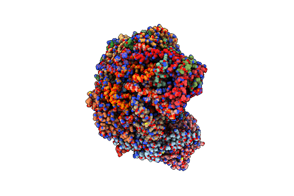 |
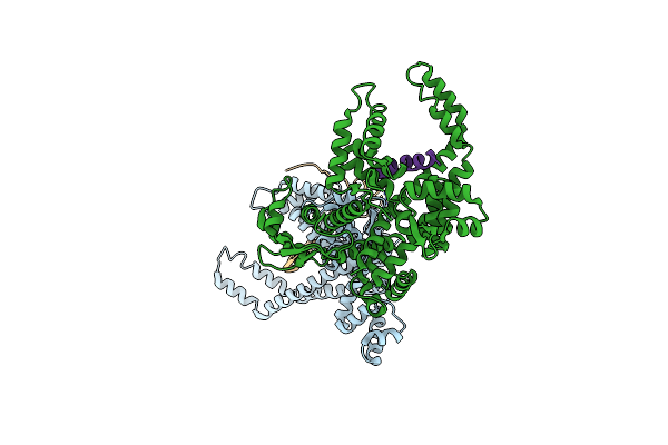
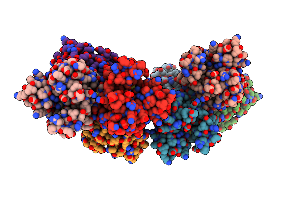
Ssu(Head) Structure Derived From The Ssu Sample Of The Mitoribosome From T. Gondii.
Organism: Toxoplasma gondii
Method: ELECTRON MICROSCOPY Release Date: 2024-12-11 Classification: RIBOSOME |
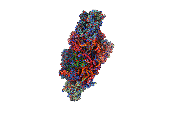 |
Ssu(Body) Structure Derived From The Ssu Sample Of The Mitoribosome From T. Gondii.
Organism: Toxoplasma gondii
Method: ELECTRON MICROSCOPY Release Date: 2024-12-11 Classification: RIBOSOME |
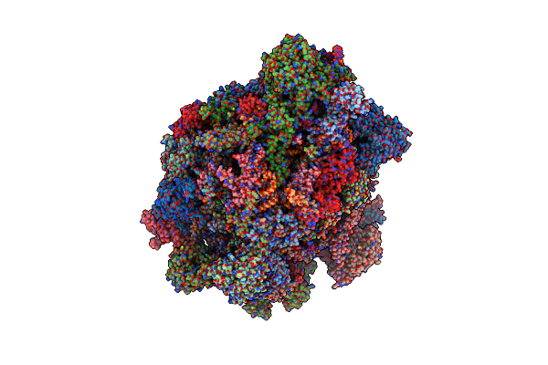 |
Lsu Structure Derived From The Lsu Sample Of The Mitoribosome From T. Gondii.
Organism: Toxoplasma gondii
Method: ELECTRON MICROSCOPY Release Date: 2024-12-11 Classification: RIBOSOME |
 |
Organism: Oryctolagus cuniculus
Method: ELECTRON MICROSCOPY Release Date: 2024-03-20 Classification: TRANSLATION Ligands: MG |
 |
Organism: Oryctolagus cuniculus
Method: ELECTRON MICROSCOPY Release Date: 2024-03-20 Classification: TRANSLATION Ligands: MG |
 |
Domain Characterization Of The Wd Protein Pwp2 And Their Relevance In Ribosome Biogenesis
Organism: Saccharomyces cerevisiae
Method: X-RAY DIFFRACTION Resolution:2.54 Å Release Date: 2017-06-21 Classification: BIOSYNTHETIC PROTEIN Ligands: SO4 |
 |
Organism: Homo sapiens
Method: X-RAY DIFFRACTION Resolution:7.00 Å Release Date: 2015-10-07 Classification: REPLICATION |
 |
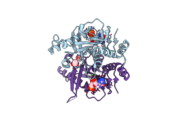
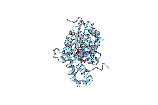
Orthorhombic Crystal Form C222 Of The Aquifex Aeolicus Nucleoside Diphosphate Kinase
Organism: Aquifex aeolicus
Method: X-RAY DIFFRACTION Resolution:1.47 Å Release Date: 2012-03-14 Classification: TRANSFERASE Ligands: SO4 |
 |
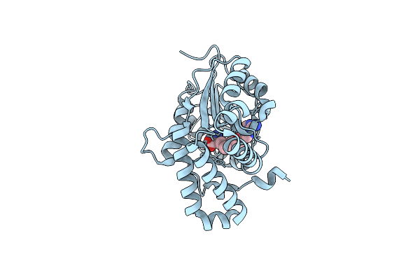
Orthorhombic Crystal Form P21212 Of The Aquifex Aeolicus Nucleoside Diphosphate Kinase
Organism: Aquifex aeolicus
Method: X-RAY DIFFRACTION Resolution:1.37 Å Release Date: 2012-03-14 Classification: TRANSFERASE Ligands: GOL, SO4 |
 |
Hexagonal Form P6122 Of The Aquifex Aeolicus Nucleoside Diphosphate Kinase (First Stage Of Radiation Damage)
Organism: Aquifex aeolicus
Method: X-RAY DIFFRACTION Resolution:2.30 Å Release Date: 2012-03-14 Classification: TRANSFERASE |
 |
Hexagonal Form P6122 Of The Aquifex Aeolicus Nucleoside Diphosphate Kinase (Final Stage Of Radiation Damage)
Organism: Aquifex aeolicus
Method: X-RAY DIFFRACTION Resolution:2.30 Å Release Date: 2012-03-14 Classification: TRANSFERASE |
 |
Hexagonal Crystal Form P61 Of The Aquifex Aeolicus Nucleoside Diphosphate Kinase
Organism: Aquifex aeolicus
Method: X-RAY DIFFRACTION Resolution:2.10 Å Release Date: 2012-02-29 Classification: TRANSFERASE |
 |
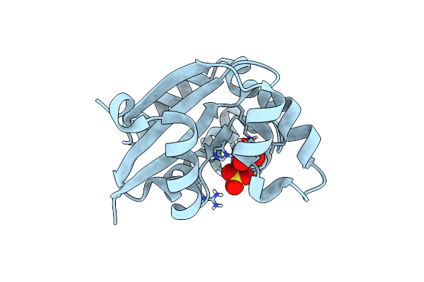
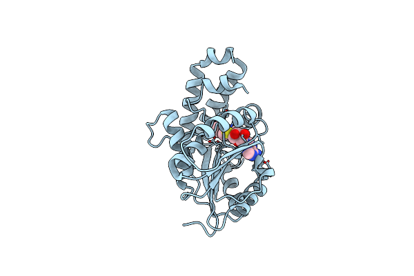
Saccharomyces Cerevisiae Hypoxanthine-Guanine Phosphoribosyltransferase In Complex With Gmp (Monoclinic Crystal Form)
Organism: Saccharomyces cerevisiae
Method: X-RAY DIFFRACTION Resolution:1.80 Å Release Date: 2011-05-04 Classification: TRANSFERASE Ligands: ACT, 5GP, MG |
 |
Saccharomyces Cerevisiae Hypoxanthine-Guanine Phosphoribosyltransferase In Complex With Gmp (Guanosine 5'- Monophosphate) (Tetragonal Crystal Form)
Organism: Saccharomyces cerevisiae
Method: X-RAY DIFFRACTION Resolution:2.30 Å Release Date: 2009-11-17 Classification: TRANSFERASE Ligands: 5GP, SO4, MG |
 |
Saccharomyces Cerevisiae Hypoxanthine-Guanine Phosphoribosyltransferase In Complex With Gmp (Guanosine 5'- Monophosphate) (Orthorhombic Crystal Form)
Organism: Saccharomyces cerevisiae
Method: X-RAY DIFFRACTION Resolution:3.45 Å Release Date: 2009-11-17 Classification: TRANSFERASE Ligands: 5GP, SO4 |
 |
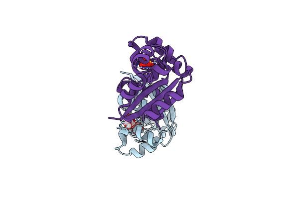
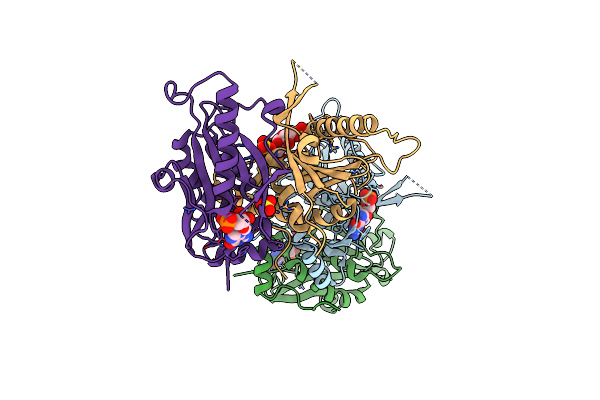
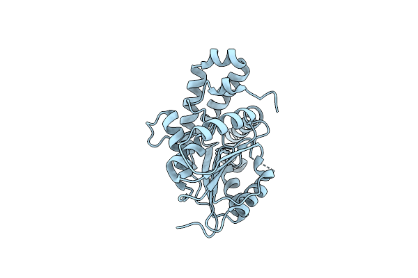
Crystal Structure Of Hma (Mmaa4) From Mycobacterium Tuberculosis Complexed With S-Adenosylhomocysteine
Organism: Mycobacterium tuberculosis
Method: X-RAY DIFFRACTION Resolution:2.20 Å Release Date: 2009-05-12 Classification: TRANSFERASE Ligands: SAH |
 |
Crystal Structure Of Hma (Mmaa4) From Mycobacterium Tuberculosis Complexed With Sinefungin
Organism: Mycobacterium tuberculosis
Method: X-RAY DIFFRACTION Resolution:2.30 Å Release Date: 2009-05-12 Classification: TRANSFERASE Ligands: SFG |
 |
Crystal Structure Of Hma (Mmaa4) From Mycobacterium Tuberculosis Complexed With S-Adenosyl-N-Decyl-Aminoethyl (Sadae)
Organism: Mycobacterium tuberculosis
Method: X-RAY DIFFRACTION Resolution:2.35 Å Release Date: 2009-05-12 Classification: TRANSFERASE Ligands: B32 |
 |
Organism: Mycobacterium tuberculosis
Method: X-RAY DIFFRACTION Resolution:2.10 Å Release Date: 2006-01-17 Classification: TRANSFERASE |
 |
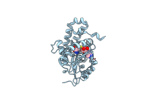
Crystal Structure Of Hma (Mmaa4) From Mycobacterium Tuberculosis Complexed With S-Adenosylmethionine
Organism: Mycobacterium tuberculosis
Method: X-RAY DIFFRACTION Resolution:2.00 Å Release Date: 2006-01-17 Classification: TRANSFERASE Ligands: SAM |