
Search Count: 25
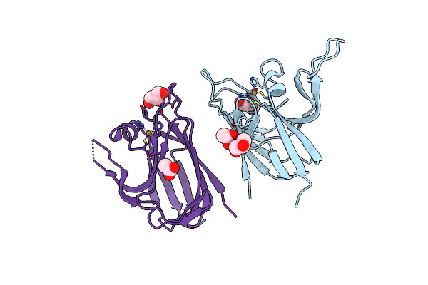 |
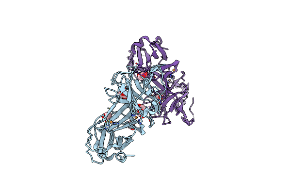
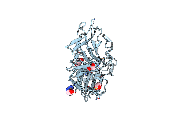
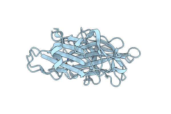
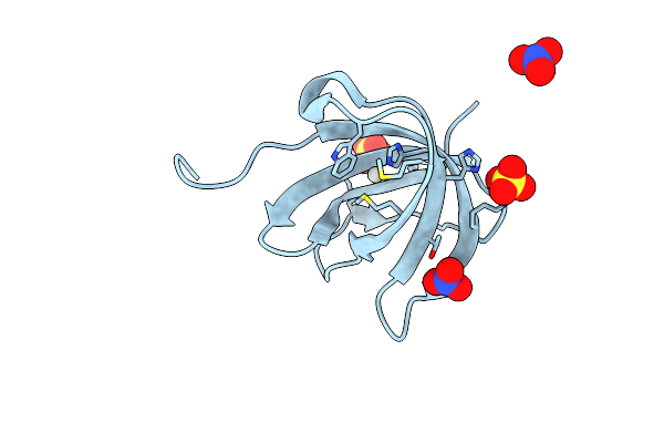
Crystal Structure Of The Cupredoxin Acop From Acidithiobacillus Ferrooxidans, Reduced Form
Organism: Acidithiobacillus ferrooxidans
Method: X-RAY DIFFRACTION Resolution:1.65 Å Release Date: 2023-09-13 Classification: METAL BINDING PROTEIN Ligands: CU1, GOL, ACT |
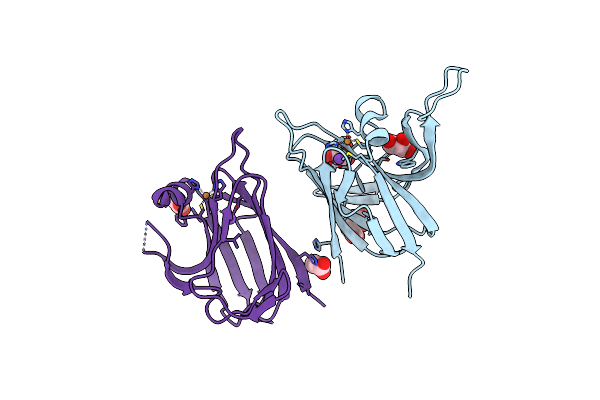 |
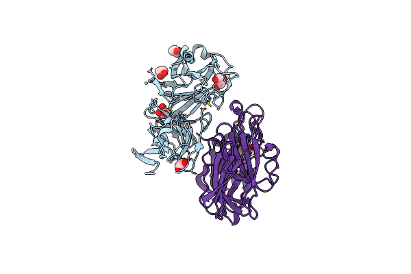
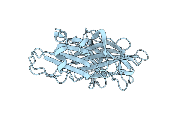
Crystal Structure Of The Cupredoxin Acop From Acidithiobacillus Ferrooxidans, Oxidized Form
Organism: Acidithiobacillus ferrooxidans
Method: X-RAY DIFFRACTION Resolution:1.70 Å Release Date: 2023-09-13 Classification: METAL BINDING PROTEIN Ligands: CU, ACT, NA, CL, GOL |
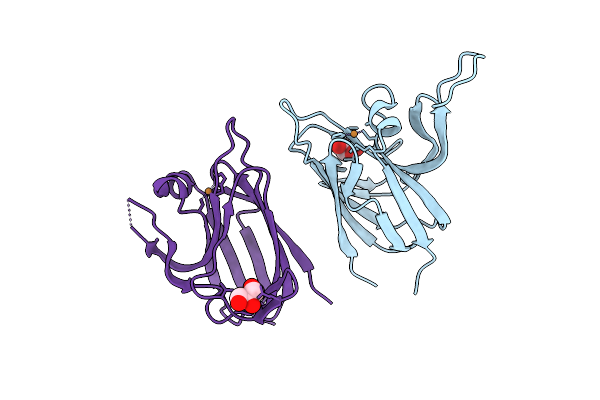 |
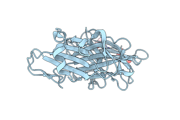
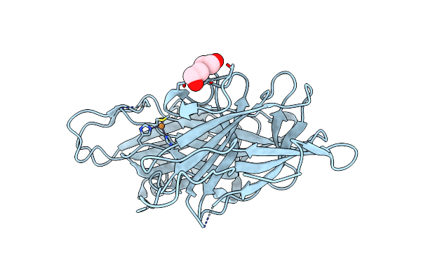
Crystal Structure Of The Cupredoxin Acop From Acidithiobacillus Ferrooxidans, H166A Mutant
Organism: Acidithiobacillus ferrooxidans
Method: X-RAY DIFFRACTION Resolution:2.10 Å Release Date: 2023-09-13 Classification: METAL BINDING PROTEIN Ligands: CU1, GOL |
 |
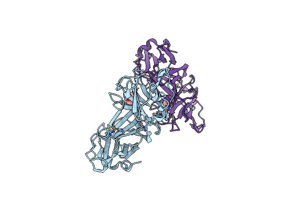
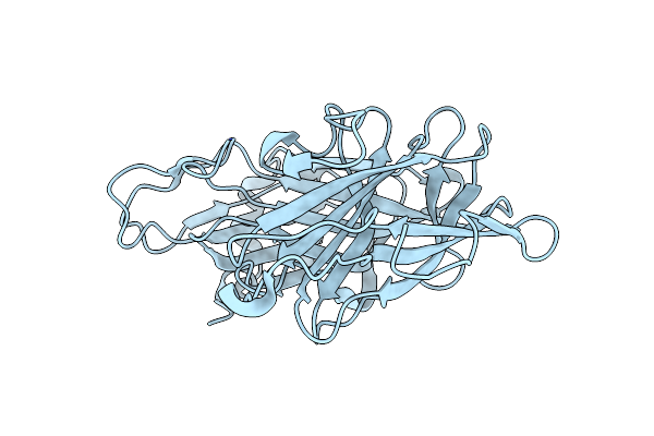
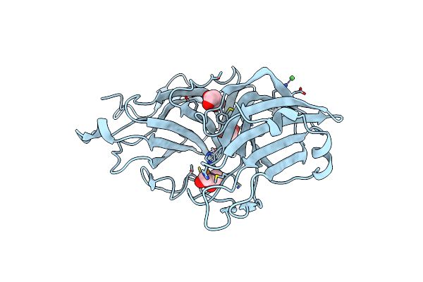
Crystal Structure Of The Cupredoxin Acop From Acidithiobacillus Ferrooxidans, M171A Mutant
Organism: Acidithiobacillus ferrooxidans
Method: X-RAY DIFFRACTION Resolution:1.82 Å Release Date: 2023-09-13 Classification: METAL BINDING PROTEIN Ligands: CU, ACT, GOL |
 |
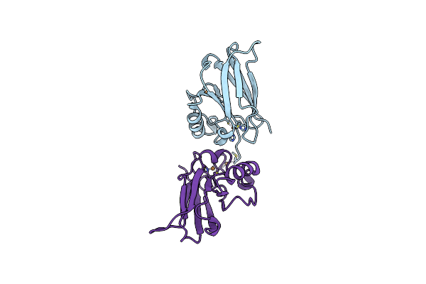
Organism: Rattus norvegicus
Method: X-RAY DIFFRACTION Resolution:2.80 Å Release Date: 2023-03-22 Classification: OXIDOREDUCTASE Ligands: CU, GOL |
 |
Organism: Rattus norvegicus
Method: X-RAY DIFFRACTION Resolution:2.05 Å Release Date: 2023-03-22 Classification: OXIDOREDUCTASE Ligands: CU, GOL |
 |
Organism: Rattus norvegicus
Method: X-RAY DIFFRACTION Resolution:2.80 Å Release Date: 2023-03-22 Classification: OXIDOREDUCTASE Ligands: CU, GOL |
 |
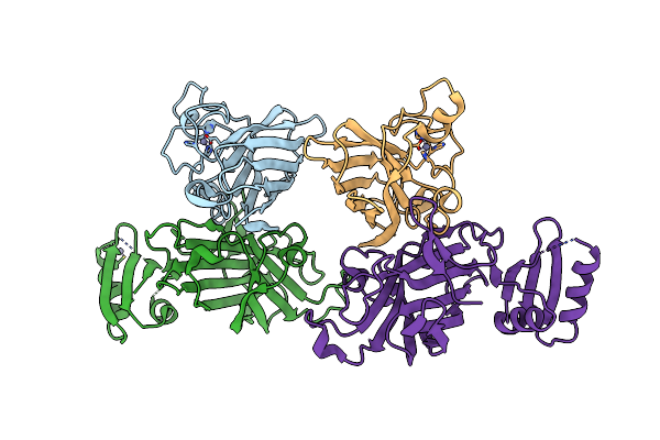
Crystal Structure Of H107A Peptidylglycine Alpha-Hydroxylating Monooxygenase (Phm) In Complex With Citrate
Organism: Rattus norvegicus
Method: X-RAY DIFFRACTION Resolution:2.30 Å Release Date: 2018-07-18 Classification: OXIDOREDUCTASE Ligands: CU, GOL, NI, FLC |
 |
Crystal Structure Of Apo Wild Type Peptidylglycine Alpha-Hydroxylating Monooxygenase (Phm)
Organism: Rattus norvegicus
Method: X-RAY DIFFRACTION Resolution:1.79 Å Release Date: 2018-07-18 Classification: OXIDOREDUCTASE Ligands: PEG, GOL |
 |
Crystal Structure Of Apo Wild Type Peptidylglycine Alpha-Hydroxylating Monooxygenase (Phm) Soaked With Peptide (Peptide Not Observed)
Organism: Rattus norvegicus
Method: X-RAY DIFFRACTION Resolution:2.40 Å Release Date: 2018-07-18 Classification: OXIDOREDUCTASE |
 |
Crystal Structure Of H108A Peptidylglycine Alpha-Hydroxylating Monooxygenase (Phm) In Complex With Citrate
Organism: Rattus norvegicus
Method: X-RAY DIFFRACTION Resolution:2.59 Å Release Date: 2018-07-18 Classification: OXIDOREDUCTASE Ligands: CU, FLC, GOL |
 |
Crystal Structure Of H107A-Peptidylglycine Alpha-Hydroxylating Monooxygenase (Phm) Mutant (No Cuh Bound)
Organism: Rattus norvegicus
Method: X-RAY DIFFRACTION Resolution:3.50 Å Release Date: 2018-07-18 Classification: OXIDOREDUCTASE Ligands: CU, AZI, GOL |
 |
Organism: Rattus norvegicus
Method: X-RAY DIFFRACTION Resolution:2.48 Å Release Date: 2018-07-18 Classification: OXIDOREDUCTASE Ligands: CU |
 |
Crystal Structure Of H172A-Peptidylglycine Alpha-Hydroxylating Monooxygenase (Phm) Mutant Soaked With Peptide (No Cuh Bound, No Peptide Bound)
Organism: Rattus norvegicus
Method: X-RAY DIFFRACTION Resolution:2.05 Å Release Date: 2018-07-18 Classification: OXIDOREDUCTASE Ligands: CU, PEG |
 |
Crystal Structure Of H108A Peptidylglycine Alpha-Hydroxylating Monooxygenase (Phm)
Organism: Rattus norvegicus
Method: X-RAY DIFFRACTION Resolution:2.98 Å Release Date: 2018-07-18 Classification: OXIDOREDUCTASE Ligands: CU, NI, GOL |
 |
Crystal Structure Of H108A Peptidylglycine Alpha-Hydroxylating Monooxygenase (Phm) Soaked With Peptide
Organism: Rattus norvegicus
Method: X-RAY DIFFRACTION Resolution:2.60 Å Release Date: 2018-07-18 Classification: OXIDOREDUCTASE Ligands: CU |
 |
Organism: Pseudomonas aeruginosa
Method: X-RAY DIFFRACTION Resolution:2.40 Å Release Date: 2017-08-16 Classification: OXIDOREDUCTASE Ligands: CU |
 |
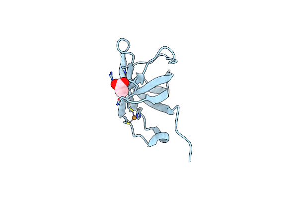
Copper-Zinc Superoxide Dismutase Is Activated Through A Sulfenic Acid Intermediate At A Copper-Ion Entry Site
Organism: Homo sapiens, Saccharomyces cerevisiae (strain atcc 204508 / s288c)
Method: X-RAY DIFFRACTION Resolution:2.35 Å Release Date: 2017-05-31 Classification: oxidoreductase/chaperone Ligands: ZN |
 |
Organism: Escherichia coli
Method: X-RAY DIFFRACTION Resolution:1.00 Å Release Date: 2009-07-07 Classification: METAL BINDING PROTEIN Ligands: CU, ACT |
 |
Organism: Escherichia coli str. k12 substr.
Method: X-RAY DIFFRACTION Resolution:1.00 Å Release Date: 2007-10-02 Classification: METAL BINDING PROTEIN Ligands: NO3, SO4, AG |