Planned Maintenance: Some services may turn out to be unavailable from 15th January, 2026 to 16th January, 2026. We apologize for the inconvenience!

Planned Maintenance: Some services may turn out to be unavailable from 15th January, 2026 to 16th January, 2026. We apologize for the inconvenience!
 |
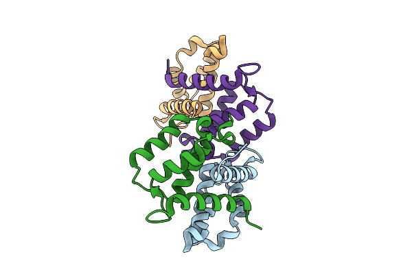
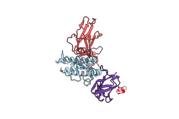
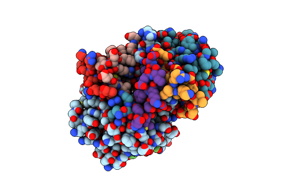
Organism: Danio rerio
Method: X-RAY DIFFRACTION Release Date: 2025-12-03 Classification: SIGNALING PROTEIN |
 |
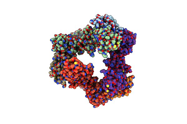
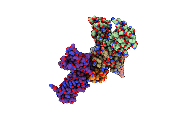
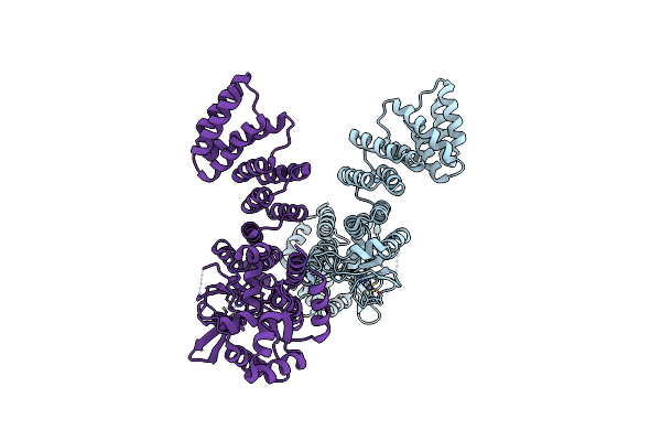
Organism: Danio rerio
Method: X-RAY DIFFRACTION Release Date: 2025-12-03 Classification: SIGNALING PROTEIN Ligands: MG |
 |
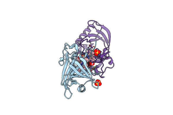
Organism: Danio rerio
Method: X-RAY DIFFRACTION Release Date: 2025-12-03 Classification: SIGNALING PROTEIN |
 |
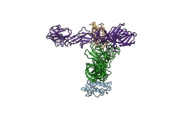
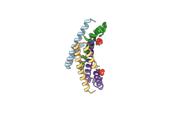
Crystal Structure For The Fniii Module Of Mouse Lep-R In Complex With The Anti-Lep-R Nanobody Vhh-4.80
Organism: Mus musculus, Lama glama
Method: X-RAY DIFFRACTION Resolution:1.75 Å Release Date: 2023-04-05 Classification: CYTOKINE Ligands: NAG, SO4 |
 |
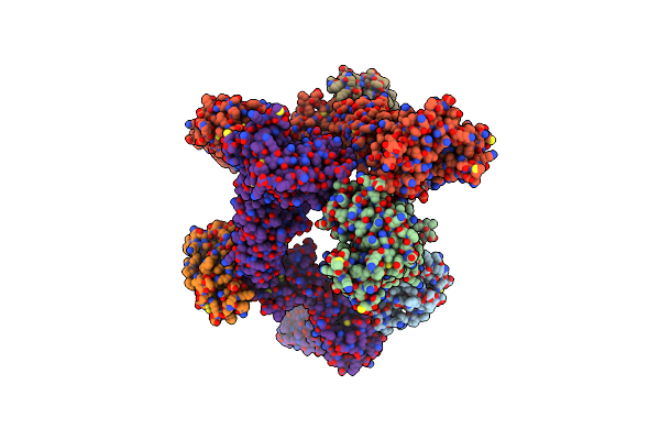
Cryo-Em Structure For Mouse Leptin In Complex With The Mouse Lep-R Ectodomain (1:2 Mlep:Mlepr Model).
Organism: Mus musculus
Method: ELECTRON MICROSCOPY Release Date: 2023-04-05 Classification: CYTOKINE |
 |
Organism: Mus musculus
Method: ELECTRON MICROSCOPY Release Date: 2023-04-05 Classification: CYTOKINE Ligands: NI |
 |
Cryo-Em Structure For A 3:3 Complex Between Mouse Leptin And The Mouse Lep-R Ectodomain (Local Refinement)
Organism: Mus musculus
Method: ELECTRON MICROSCOPY Release Date: 2023-04-05 Classification: CYTOKINE Ligands: NI |
 |
Human Leptin In Complex With The Human Lep-R Ectodomain Fused To A C-Terminal Trimeric Isoleucine Gcn4 Zipper (2:2 Model)
Organism: Homo sapiens
Method: ELECTRON MICROSCOPY Release Date: 2023-04-05 Classification: CYTOKINE |
 |
Human Leptin In Complex With The Human Lep-R Ectodomain Fused To A C-Terminal Trimeric Isoleucine Gcn4 Zipper (Closed 3:3 Model)
Organism: Homo sapiens
Method: ELECTRON MICROSCOPY Release Date: 2023-04-05 Classification: CYTOKINE |
 |
Human Leptin In Complex With The Human Lep-R Ectodomain Fused To A C-Terminal Trimeric Isoleucine Gcn4 Zipper (Open 3:3 Model).
Organism: Homo sapiens
Method: ELECTRON MICROSCOPY Release Date: 2023-04-05 Classification: CYTOKINE |
 |
Cryo-Em Structure For The Mouse Lepr-Crh2:Leptin:Lepr-Ig Complex Following Symmetry Expansion In Combination With Local Refinement
Organism: Mus musculus
Method: ELECTRON MICROSCOPY Release Date: 2023-04-05 Classification: CYTOKINE Ligands: NAG |
 |
Crystal Structure Of The Mouse Leptin:Lepr-Crh2 Encounter Complex To 1.95 A Resolution.
Organism: Mus musculus
Method: X-RAY DIFFRACTION Resolution:1.94 Å Release Date: 2023-03-22 Classification: CYTOKINE Ligands: CA, PEG |
 |
Crystal Structure Of The Human Leptin:Lepr-Crh2 Encounter Complex To 3.6 A Resolution.
Organism: Homo sapiens
Method: X-RAY DIFFRACTION Resolution:3.62 Å Release Date: 2023-03-22 Classification: CYTOKINE |
 |
Crystal Structure Of The Mouse Leptin:Lepr-Igcrh2 Complex To 2.95 A Resolution.
Organism: Mus musculus
Method: X-RAY DIFFRACTION Resolution:2.95 Å Release Date: 2023-03-22 Classification: CYTOKINE Ligands: NAG |
 |
Organism: Mus musculus
Method: X-RAY DIFFRACTION Resolution:2.80 Å Release Date: 2022-01-26 Classification: APOPTOSIS Ligands: HEM, SO4 |
 |
Het-N2 - De Novo Designed Three-Helix Heterodimer With Cysteine At The N2 Position Of The Alpha-Helix
Organism: Synthetic construct
Method: X-RAY DIFFRACTION Resolution:2.33 Å Release Date: 2021-03-17 Classification: DE NOVO PROTEIN Ligands: CD, NA |
 |
Het-Ncap - De Novo Designed Three-Helix Heterodimer With Cysteine At The Ncap Position Of The Alpha-Helix
Organism: Synthetic construct
Method: X-RAY DIFFRACTION Resolution:1.45 Å Release Date: 2021-03-17 Classification: DE NOVO PROTEIN Ligands: SO4, ZN |
 |
Het-N2-So3- - De Novo Designed Three-Helix Heterodimer With Cysteine S-Sulfate At The N2 Position Of The Alpha-Helix
Organism: Synthetic construct
Method: X-RAY DIFFRACTION Resolution:1.50 Å Release Date: 2021-03-17 Classification: DE NOVO PROTEIN Ligands: SO4 |
 |
Tctp Contains A Bh3-Like Domain, Which Instead Of Inhibiting, Activates Bcl-Xl
Organism: Homo sapiens
Method: X-RAY DIFFRACTION Resolution:2.10 Å Release Date: 2016-02-10 Classification: APOPTOSIS Ligands: BCT, NA |
 |
Organism: Arabidopsis thaliana
Method: X-RAY DIFFRACTION Resolution:3.05 Å Release Date: 2016-01-27 Classification: HYDROLASE Ligands: ZN |