
Search Count: 52
 |
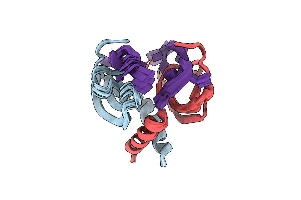
Organism: Salmonella enterica subsp. enterica serovar typhimurium
Method: SOLUTION NMR Release Date: 2023-07-05 Classification: CHAPERONE |
 |
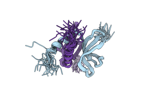
Organism: Salmonella enterica subsp. enterica serovar typhimurium
Method: SOLUTION NMR Release Date: 2023-07-05 Classification: CHAPERONE |
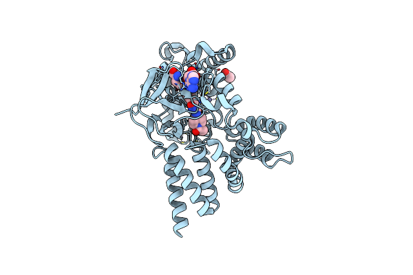 |
Crystal Structure Of Smyd3 Conjugate With Piperidine-Based Covalent Inhibitor Em127
Organism: Homo sapiens
Method: X-RAY DIFFRACTION Resolution:1.55 Å Release Date: 2022-07-27 Classification: ONCOPROTEIN Ligands: SAM, QON, ACT, ZN |
 |
Crystal Structure Of Smyd3 With Diperodon S Enantiomer Bound To Allosteric Site
Organism: Homo sapiens
Method: X-RAY DIFFRACTION Resolution:1.61 Å Release Date: 2021-03-03 Classification: ONCOPROTEIN Ligands: SAM, QKT, ZN, ACT, GOL |
 |
Crystal Structure Of Smyd3 With Diperodon R Enantiomer Bound To Allosteric Site
Organism: Homo sapiens
Method: X-RAY DIFFRACTION Resolution:1.93 Å Release Date: 2021-01-13 Classification: ONCOPROTEIN Ligands: SAM, GOL, POW, ZN |
 |
2.6 Angstrom Resolution Crystal Structure Of Stage Ii Sporulation Protein D (Spoiid) From Clostridium Difficile In Complex With Triacetylchitotriose
Organism: Peptoclostridium difficile (strain 630)
Method: X-RAY DIFFRACTION Resolution:2.60 Å Release Date: 2016-05-25 Classification: HYDROLASE Ligands: ZN, NA, CL, GOL |
 |
1.7 Angstrom Resolution Crystal Structure Of Putative Nucleoside Diphosphate Kinase From Toxoplasma Gondii With Tyrosine Of Tag Bound To Active Site
Organism: Toxoplasma gondii me49
Method: X-RAY DIFFRACTION Resolution:1.70 Å Release Date: 2015-06-24 Classification: TRANSFERASE Ligands: BCT, PEG |
 |
2.1 Angstrom Crystal Structure Of Stage Ii Sporulation Protein D From Bacillus Anthracis
Organism: Bacillus anthracis
Method: X-RAY DIFFRACTION Resolution:2.10 Å Release Date: 2014-12-17 Classification: VIRAL PROTEIN |
 |
1.7 Angstrom Crystal Structure Of Leukotoxin Lukd From Staphylococcus Aureus.
Organism: Staphylococcus aureus subsp. aureus
Method: X-RAY DIFFRACTION Resolution:1.70 Å Release Date: 2014-05-07 Classification: TOXIN Ligands: BTB |
 |
2.6 Angstrom Crystal Structure Of Putative Phosphoglycerate Mutase 1 From Toxoplasma Gondii
Organism: Toxoplasma gondii
Method: X-RAY DIFFRACTION Resolution:2.60 Å Release Date: 2014-01-22 Classification: ISOMERASE |
 |
2.4 Angstrom Resolution Crystal Structure Of Putative Nucleoside Diphosphate Kinase From Toxoplasma Gondii.
Organism: Toxoplasma gondii me49
Method: X-RAY DIFFRACTION Resolution:2.40 Å Release Date: 2013-12-25 Classification: TRANSFERASE Ligands: SO4 |
 |
2.05 Angstrom Crystal Structure Of Ribulose-Phosphate 3-Epimerase From Toxoplasma Gondii.
Organism: Toxoplasma gondii
Method: X-RAY DIFFRACTION Resolution:2.05 Å Release Date: 2013-12-18 Classification: ISOMERASE Ligands: CL, ZN, SO4 |
 |
2.60 Angstrom Resolution Crystal Structure Of Putative Ribose 5-Phosphate Isomerase From Toxoplasma Gondii Me49 In Complex With Dl-Malic Acid
Organism: Toxoplasma gondii
Method: X-RAY DIFFRACTION Resolution:2.60 Å Release Date: 2013-12-04 Classification: ISOMERASE Ligands: CL, MLT |
 |
Crystal Structure Of A Putative Ornithine Aminotransferase From Toxoplasma Gondii Me49 In Complex With Pyrodoxal-5'-Phosphate
Organism: Toxoplasma gondii
Method: X-RAY DIFFRACTION Release Date: 2013-12-04 Classification: TRANSFERASE Ligands: PLP, BME, ACT, BTB, PEG |
 |
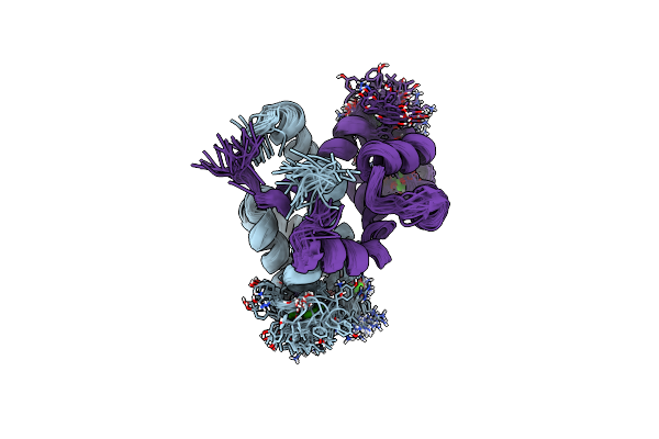
Solution Nmr Structure Of Ydbc:Dt19G1 Complex. Northeast Structural Genomics Consortium (Nesg) Target Kr150
Organism: Lactococcus lactis subsp. lactis
Method: SOLUTION NMR Release Date: 2012-06-20 Classification: TRANSCRIPTION, DNA BINDING PROTEIN/DNA |
 |
Solution Nmr Structure Of Apo Ydbc From Lactococcus Lactis, Northeast Structural Genomics Consortium (Nesg) Target Kr150
Organism: Lactococcus lactis subsp. lactis il1403
Method: SOLUTION NMR Release Date: 2012-06-06 Classification: Structural Genomics, Unknown Function |
 |
Crystal Structure Of Leukotoxin (Luke) From Staphylococcus Aureus Subsp. Aureus Col.
Organism: Staphylococcus aureus subsp. aureus nctc 8325
Method: X-RAY DIFFRACTION Resolution:3.20 Å Release Date: 2011-05-04 Classification: TOXIN Ligands: PGE, CL |
 |
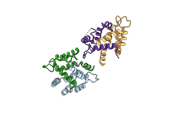
Organism: Homo sapiens
Method: SOLUTION NMR Release Date: 2010-11-03 Classification: METAL BINDING PROTEIN |
 |
Organism: Homo sapiens
Method: SOLUTION NMR Release Date: 2010-11-03 Classification: METAL BINDING PROTEIN Ligands: CA |
 |
Organism: Homo sapiens
Method: X-RAY DIFFRACTION Resolution:2.10 Å Release Date: 2010-11-03 Classification: METAL BINDING PROTEIN |