
Search Count: 14
 |
Crystal Structure Of The Novel Complex Formed Between Zinc 2-Glycoprotein (Zag) And Prolactin Inducible Protein (Pip) From Human Seminal Plasma
Organism: Homo sapiens
Method: X-RAY DIFFRACTION Resolution:3.23 Å Release Date: 2008-10-28 Classification: CELL ADHESION Ligands: CO3, P6G |
 |
Crystal Structure Of The Buffalo Secretory Signalling Glycoprotein At 2.8 A Resolution
Organism: Bubalus bubalis
Method: X-RAY DIFFRACTION Resolution:2.80 Å Release Date: 2007-01-02 Classification: SIGNALING PROTEIN |
 |
Crystal Structure Of A Novel Disintegrin From Saw-Scaled Viper At 3.2 A Resolution
Organism: Echis carinatus
Method: X-RAY DIFFRACTION Resolution:3.20 Å Release Date: 2005-04-19 Classification: PROTEIN BINDING |
 |
Crystal Structure Of Himalayan Mistletoe Rip Reveals The Presence Of A Natural Inhibitor And A New Functionally Active Sugar-Binding Site
Organism: Viscum album
Method: X-RAY DIFFRACTION Resolution:2.80 Å Release Date: 2005-03-08 Classification: HYDROLASE Ligands: NAG, P6C, GAL |
 |
Crystal Structure Of A Ribosome Inactivating Protein In Its Naturally Inhibited Form
Organism: Viscum album
Method: X-RAY DIFFRACTION Resolution:2.80 Å Release Date: 2004-07-13 Classification: TOXIN Ligands: NAG, P6C, GAL |
 |
Crystal Structure Of A Buffalo Signaling Glycoprotein (Spb-40) Secreted During Involution
Organism: Bubalus bubalis
Method: X-RAY DIFFRACTION Resolution:2.90 Å Release Date: 2004-07-13 Classification: SIGNALING PROTEIN |
 |
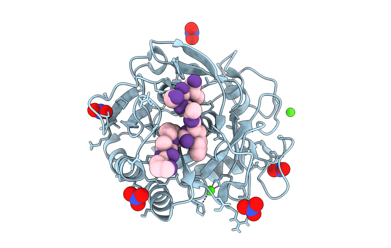
Crystal Structure Of Russells Viper Phospholipase A2 In Complex With A Specifically Designed Tetrapeptide Ala-Ile-Arg-Ser At 1.1 A Resolution
Organism: Daboia russellii pulchella
Method: X-RAY DIFFRACTION Resolution:1.10 Å Release Date: 2004-06-22 Classification: HYDROLASE Ligands: SO4 |
 |
Crystal Structure Of Schistatin, A Disintegrin Homodimer From Saw-Scaled Viper (Echis Carinatus) At 2.5 A Resolution
Organism: Echis carinatus
Method: X-RAY DIFFRACTION Resolution:2.50 Å Release Date: 2004-06-16 Classification: PROTEIN BINDING |
 |
Organism: Echis carinatus
Method: X-RAY DIFFRACTION Resolution:1.90 Å Release Date: 2004-06-15 Classification: PROTEIN BINDING |
 |
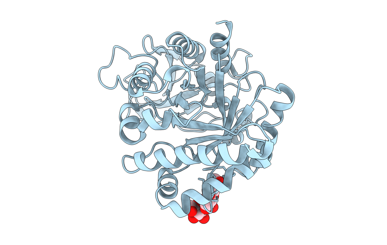
Crystal Structure Of A Complex Formed Between Organic Solvent Treated Bovine Alpha-Chymotrypsin And Its Autocatalytically Produced Highly Potent 14-Residue Peptide At 2.2 Resolution
Organism: Bos taurus
Method: X-RAY DIFFRACTION Resolution:2.20 Å Release Date: 2004-05-18 Classification: HYDROLASE Ligands: SO4 |
 |
Structure Of A Complex Formed Between Proteinase K And A Designed Heptapeptide Inhibitor Pro-Ala-Pro-Phe-Ala-Ala-Ala At Atomic Resolution
Organism: Engyodontium album
Method: X-RAY DIFFRACTION Resolution:1.08 Å Release Date: 2004-05-18 Classification: HYDROLASE Ligands: CA, NO3 |
 |
Crystal Structure Of The Complex Of Proteinase K With A Designed Heptapeptide Inhibitor Pro-Ala-Pro-Phe-Ala-Ser-Ala At Atomic Resolution
Organism: Engyodontium album
Method: X-RAY DIFFRACTION Resolution:1.02 Å Release Date: 2004-05-18 Classification: HYDROLASE Ligands: CA, NO3 |
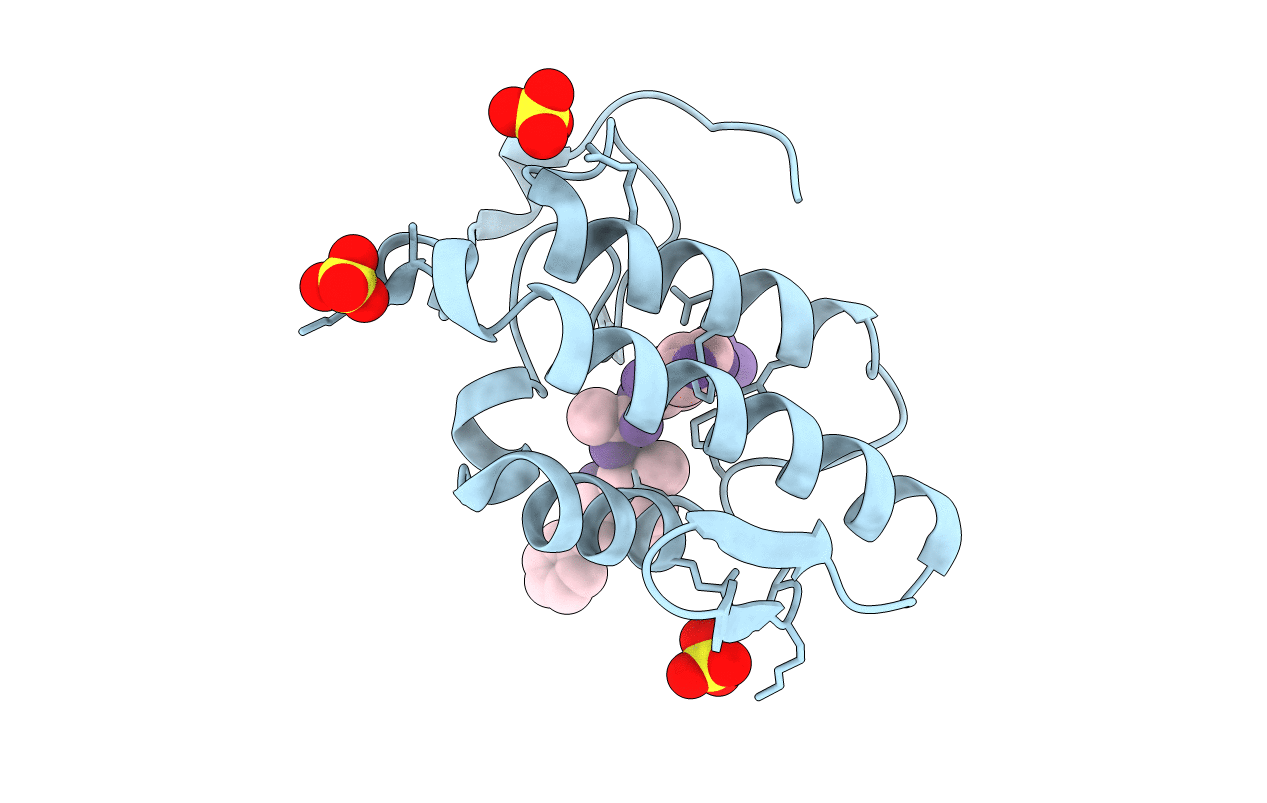 |
Design Of Specific Inhibitors Of Phopholipase A2: Crystal Structure Of The Complex Formed Between Group Ii Phopholipase A2 And A Designed Peptide Dehydro-Ile-Ala-Arg-Ser At 1.2A Resolution
Organism: Daboia russellii russellii
Method: X-RAY DIFFRACTION Resolution:1.20 Å Release Date: 2004-04-13 Classification: HYDROLASE/HYDROLASE INHIBITOR Ligands: SO4 |
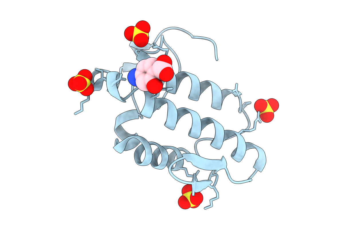 |
Crystal Structure Of A Complex Formed Between Phospholipase A2 And A Non-Specific Anti-Inflammatory Amino Salicylic Acid At 1.2 A Resolution
Organism: Daboia russellii russellii
Method: X-RAY DIFFRACTION Resolution:1.21 Å Release Date: 2004-04-13 Classification: TOXIN Ligands: SO4, BHA |