
Search Count: 25
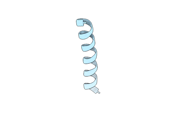 |
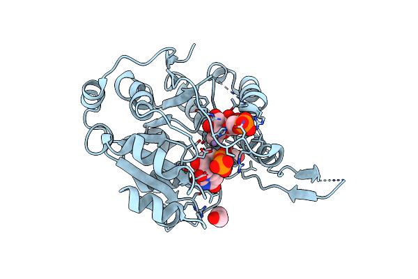
Atomic Resolution Crystal Structure Of The Hexameric Antimicrobial Peptide Magainin-2
Organism: Xenopus laevis
Method: X-RAY DIFFRACTION Resolution:1.05 Å Release Date: 2025-02-05 Classification: ANTIBIOTIC |
 |
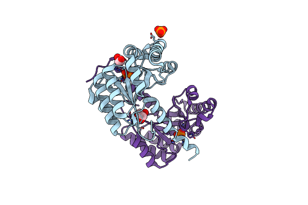
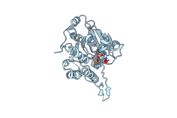
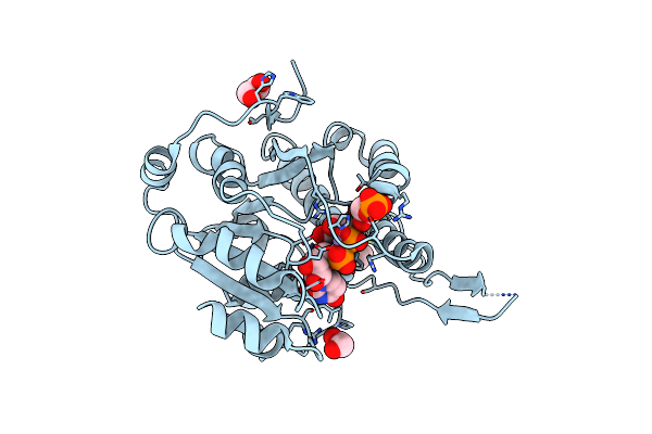
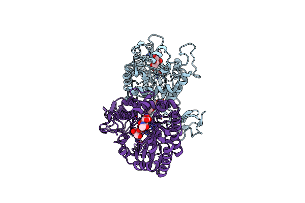
Crystal Structure Of Udp-Galactose 4-Epimerase From Pyrococcus Horikoshii With Bound Nad
Organism: Pyrococcus horikoshii
Method: X-RAY DIFFRACTION Resolution:1.90 Å Release Date: 2024-12-18 Classification: ISOMERASE Ligands: NAD |
 |
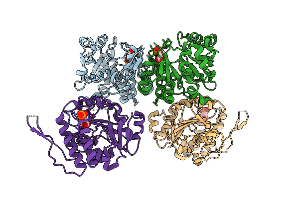
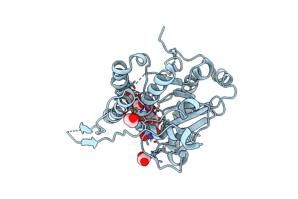
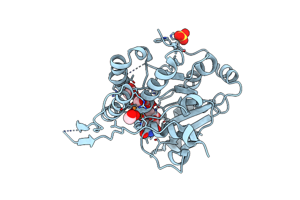
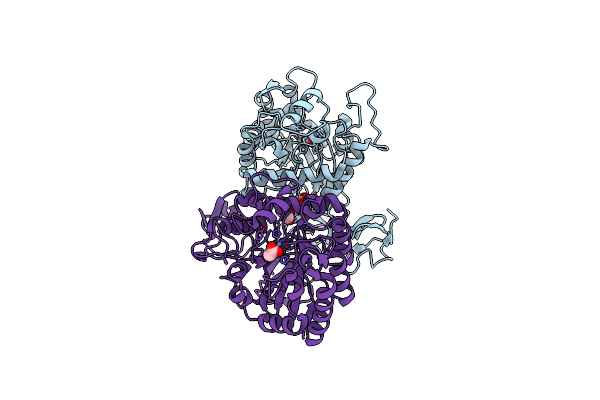
Crystal Structure Of Udp-Galactose 4-Epimerase From Pyrococcus Horikoshii With Bound Nad And Gdp-L-Fucose
Organism: Pyrococcus horikoshii
Method: X-RAY DIFFRACTION Resolution:2.40 Å Release Date: 2024-12-18 Classification: ISOMERASE Ligands: NAD, GFB |
 |
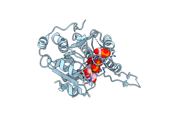
Crystal Structure Of Udp-Galactose 4-Epimerase From Pyrococcus Horikoshii Containing Y145F Mutation And With Bound Nad And Gdp-L-Fucose
Organism: Pyrococcus horikoshii
Method: X-RAY DIFFRACTION Resolution:3.10 Å Release Date: 2024-12-18 Classification: ISOMERASE Ligands: NAD, GFB |
 |
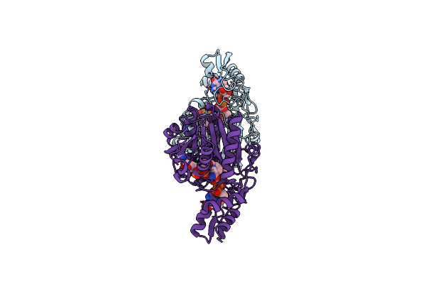
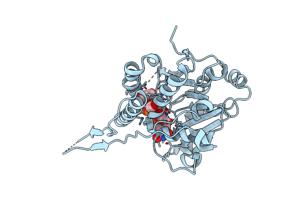
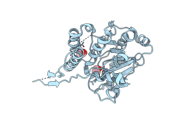
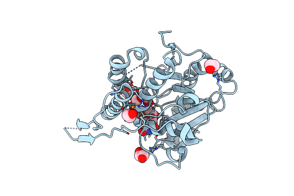
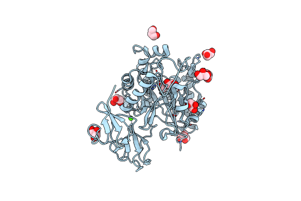
Organism: Bacillus subtilis subsp. subtilis str. 168
Method: X-RAY DIFFRACTION Resolution:1.54 Å Release Date: 2019-11-13 Classification: HYDROLASE Ligands: ZN, TLA |
 |
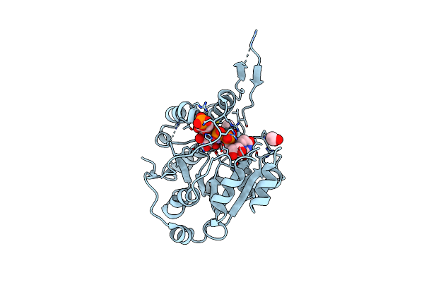
Structure Of Peptidoglycan Deacetylase Pdac From Bacillus Subtilis - Mutant D285S
Organism: Bacillus subtilis subsp. subtilis str. 168
Method: X-RAY DIFFRACTION Resolution:1.26 Å Release Date: 2019-11-13 Classification: HYDROLASE Ligands: ZN, PO4, GOL |
 |
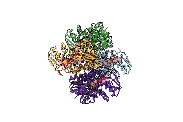
Crystal Structure Of Glucosyl-3-Phosphoglycerate Synthase From Mycobacterium Tuberculosis In Complex With Phosphoglyceric Acid (Pga) - Gpgs*Pga
Organism: Mycobacterium tuberculosis h37rv
Method: X-RAY DIFFRACTION Resolution:2.82 Å Release Date: 2017-05-24 Classification: TRANSFERASE Ligands: 3PG |
 |
Crystal Structure Of Glucosyl-3-Phosphoglycerate Synthase From Mycobacterium Tuberculosis In Complex With Mn2+ And Uridine-Diphosphate-Glucose (Udp-Glc)
Organism: Mycobacterium tuberculosis h37ra
Method: X-RAY DIFFRACTION Resolution:2.81 Å Release Date: 2017-05-24 Classification: TRANSFERASE Ligands: MN, UPG, GOL |
 |
Crystal Structure Of Glucosyl-3-Phosphoglycerate Synthase From Mycobacterium Tuberculosis In Complex With Mn2+, Uridine-Diphosphate (Udp) And Glucosyl-3-Phosphoglycerate (Gpg) - Gpgs*Gpg*Udp*Mn2+
Organism: Mycobacterium tuberculosis
Method: X-RAY DIFFRACTION Resolution:2.80 Å Release Date: 2017-05-24 Classification: TRANSFERASE Ligands: MN, UDP, EDO, XDX |
 |
Crystal Structure Of Glucosyl-3-Phosphoglycerate Synthase From Mycobacterium Tuberculosis In Complex With Mn2+, Uridine-Diphosphate (Udp) And Glucosyl-3-Phosphoglycerate (Gpg) - Gpgs*Gpg*Udp*Mn2+_2
Organism: Mycobacterium tuberculosis (strain atcc 25618 / h37rv)
Method: X-RAY DIFFRACTION Resolution:2.80 Å Release Date: 2017-05-24 Classification: TRANSFERASE Ligands: UDP, EDO, MN, XDX |
 |
Crystal Structure Of Glucosyl-3-Phosphoglycerate Synthase From Mycobacterium Tuberculosis In Complex With Uridine-Diphosphate (Udp) - Gpgs*Udp
Organism: Mycobacterium bovis af2122/97
Method: X-RAY DIFFRACTION Resolution:2.59 Å Release Date: 2017-05-24 Classification: TRANSFERASE Ligands: UDP |
 |
Crystal Structure Of Glucosyl-3-Phosphoglycerate Synthase From Mycobacterium Tuberculosis - Apo Form
Organism: Mycobacterium tuberculosis h37ra
Method: X-RAY DIFFRACTION Resolution:2.60 Å Release Date: 2016-12-21 Classification: TRANSFERASE Ligands: GOL |
 |
Crystal Structure Of Glucosyl-3-Phosphoglycerate Synthase From Mycobacterium Tuberculosis In Complex With Mn2+, Uridine-Diphosphate-Glucose (Udp-Glc) And Phosphoglyceric Acid (Pga) - Gpgs Mn2+ Udp-Glc Pga-1
Organism: Mycobacterium tuberculosis
Method: X-RAY DIFFRACTION Resolution:2.35 Å Release Date: 2015-07-15 Classification: TRANSFERASE Ligands: MN, 3PG, UPG, EDO |
 |
Organism: Mycobacterium tuberculosis (strain atcc 25618 / h37rv)
Method: X-RAY DIFFRACTION Resolution:2.27 Å Release Date: 2015-07-15 Classification: TRANSFERASE Ligands: MN, CL, 3PG, UPG, EDO |
 |
Crystal Structure Of Glucosyl-3-Phosphoglycerate Synthase From Mycobacterium Tuberculosis In Complex With Mn2+, Uridine-Diphosphate-Glucose (Udp-Glc) And 3-(Phosphonooxy)Propanoic Acid (Ppa) - Gpgs Mn2+ Udp-Glc Ppa
Organism: Mycobacterium tuberculosis (strain atcc 25618 / h37rv)
Method: X-RAY DIFFRACTION Resolution:3.23 Å Release Date: 2015-07-15 Classification: TRANSFERASE Ligands: MN, 48X, UPG, SO4, EDO |
 |
Crystal Structure Of Glucosyl-3-Phosphoglycerate Synthase From Mycobacterium Tuberculosis In Complex With Mn2+, Uridine-Diphosphate-Glucose (Udp-Glc) And Glycerol 3-Phosphate (G3P) - Gpgs Mn2+ Udp-Glc G3P
Organism: Mycobacterium tuberculosis (strain atcc 25618 / h37rv)
Method: X-RAY DIFFRACTION Resolution:2.59 Å Release Date: 2015-07-15 Classification: TRANSFERASE Ligands: MN, G3P, UPG |
 |
Crystal Structure Of Glucosyl-3-Phosphoglycerate Synthase From Mycobacterium Tuberculosis In Complex With Mn2+, Uridine-Diphosphate-Glucose (Udp-Glc) And Phosphoglyceric Acid (Pga) - Gpgs Mn2+ Udp-Glc Pga-3
Organism: Mycobacterium tuberculosis (strain atcc 25618 / h37rv)
Method: X-RAY DIFFRACTION Resolution:2.64 Å Release Date: 2015-07-15 Classification: TRANSFERASE Ligands: MN, CL, 3PG, UPG, EDO |
 |
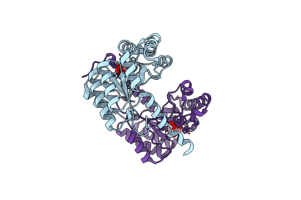
Structure Of Vibrio Cholerae Chitin De-N-Acetylase In Complex With 2-(Acetylamino)-2-Deoxy-A-D-Glucopyranose (Ndg) And Cadmium Ion
Organism: Vibrio cholerae
Method: X-RAY DIFFRACTION Resolution:1.87 Å Release Date: 2014-11-26 Classification: HYDROLASE |
 |
Structure Of Vibrio Cholerae Chitin De-N-Acetylase In Complex With Acetate Ion (Act) In P 21
Organism: Vibrio cholerae
Method: X-RAY DIFFRACTION Resolution:1.88 Å Release Date: 2014-08-13 Classification: HYDROLASE Ligands: ZN, CA, ACT, GOL |
 |
Structure Of Vibrio Cholerae Chitin De-N-Acetylase In Complex With Acetate Ion (Act) In C 2 2 21
Organism: Vibrio cholerae
Method: X-RAY DIFFRACTION Resolution:2.03 Å Release Date: 2014-08-13 Classification: HYDROLASE Ligands: ZN, CA, ACT, GOL |