
Search Count: 56
 |
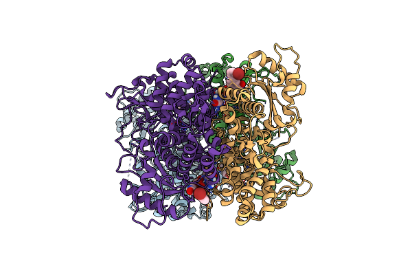
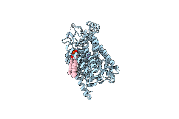
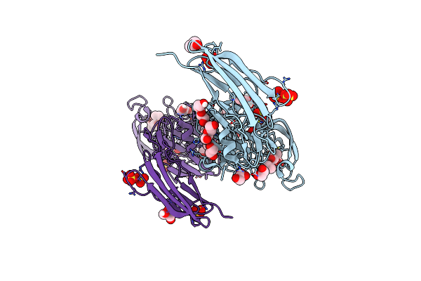
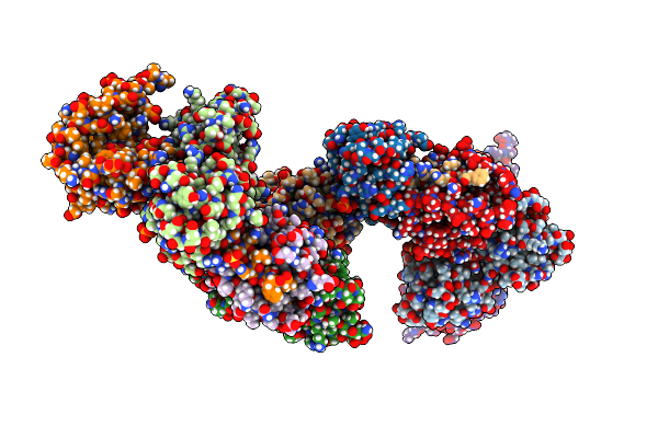
Crystal Structure Of Samhd1 Dimer Bound To An Inhibitor Obtained From High-Throughput Chemical Tethering To The Guanine Antiviral Acyclovir
Organism: Homo sapiens
Method: X-RAY DIFFRACTION Resolution:2.72 Å Release Date: 2025-03-12 Classification: HYDROLASE Ligands: A1BHL, FE |
 |
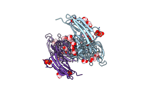
Organism: Homo sapiens
Method: X-RAY DIFFRACTION Resolution:2.46 Å Release Date: 2023-06-07 Classification: HYDROLASE Ligands: YWI, FE |
 |
Organism: Homo sapiens
Method: X-RAY DIFFRACTION Resolution:3.07 Å Release Date: 2023-06-07 Classification: HYDROLASE Ligands: FE |
 |
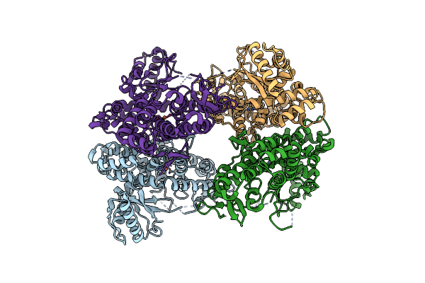
Organism: Rattus norvegicus
Method: ELECTRON MICROSCOPY Release Date: 2022-12-21 Classification: TRANSPORT PROTEIN Ligands: 3PH |
 |
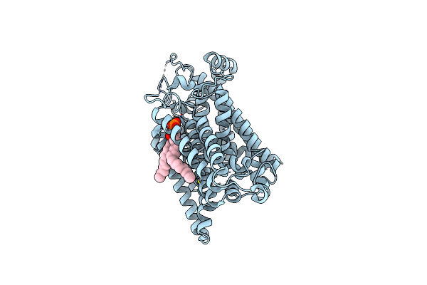
Structure Of The Sodium/Iodide Symporter (Nis) In Complex With Perrhenate And Sodium
Organism: Rattus norvegicus
Method: ELECTRON MICROSCOPY Release Date: 2022-12-21 Classification: TRANSPORT PROTEIN |
 |
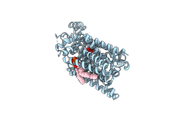
Structure Of The Sodium/Iodide Symporter (Nis) In Complex With Iodide And Sodium
Organism: Rattus norvegicus
Method: ELECTRON MICROSCOPY Release Date: 2022-12-21 Classification: TRANSPORT PROTEIN |
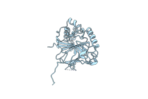 |
Organism: Schizosaccharomyces pombe
Method: X-RAY DIFFRACTION Resolution:2.02 Å Release Date: 2019-09-11 Classification: CYTOSOLIC PROTEIN |
 |
Organism: Danio rerio
Method: X-RAY DIFFRACTION Resolution:2.00 Å Release Date: 2019-03-20 Classification: SUGAR BINDING PROTEIN Ligands: MG |
 |
Crystal Structure Of Ldtmt2 (56-408) With A Panipenem Adduct At The Active Site Cysteine-354
Organism: Mycobacterium tuberculosis (strain atcc 25177 / h37ra)
Method: X-RAY DIFFRACTION Resolution:2.10 Å Release Date: 2018-04-11 Classification: TRANSFERASE Ligands: E0Y, PEG, GOL, SO4 |
 |
X-Ray Crystal Structure Of A Enzymatically Degraded Biapenem-Adduct Of L,D-Transpeptidase 2 From Mycobacterium Tuberculosis
Organism: Mycobacterium tuberculosis (strain cdc 1551 / oshkosh)
Method: X-RAY DIFFRACTION Resolution:2.18 Å Release Date: 2016-10-05 Classification: TRANSFERASE Ligands: 58U, SO4, PEG, PGE, GOL, EDO |
 |
X-Ray Crystal Structure Of L,D Transpeptidase 2 From Mycobacterium Tuberculosis
Organism: Mycobacterium tuberculosis
Method: X-RAY DIFFRACTION Resolution:2.49 Å Release Date: 2016-09-28 Classification: TRANSFERASE Ligands: SO4, GOL |
 |
X-Ray Crystal Structure Of A Tebipenem Adduct Of L,D Transpeptidase 2 From Mycobacterium Tuberculosis
Organism: Mycobacterium tuberculosis
Method: X-RAY DIFFRACTION Resolution:2.45 Å Release Date: 2016-09-28 Classification: TRANSFERASE Ligands: 58U, SO4, PEG, PGE, GOL, EDO |
 |
Construct Of Mical-1 Containing The Monooxygenase And Calponin Homology Domains
Organism: Mus musculus
Method: X-RAY DIFFRACTION Resolution:2.31 Å Release Date: 2016-01-13 Classification: OXIDOREDUCTASE Ligands: FAD, CL, PEG |
 |
Construct Of Mical-1 Containing The Monooxygenase And Calponin Homology Domains
Organism: Mus musculus
Method: X-RAY DIFFRACTION Resolution:2.88 Å Release Date: 2015-12-23 Classification: OXIDOREDUCTASE Ligands: PEG, FAD |
 |
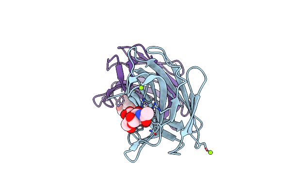
Structural And Biochemical Characterization Of A Non-Functionally Redundant M. Tuberculosis (3,3) L,D-Transpeptidase, Ldtmt5.
Organism: Mycobacterium tuberculosis (strain atcc 25177 / h37ra)
Method: X-RAY DIFFRACTION Resolution:1.98 Å Release Date: 2015-09-02 Classification: TRANSFERASE Ligands: PO4, PEG, PGE, ACE |
 |
Structure Of M. Tuberculosis (3,3) L,D-Transpeptidase, Ldtmt5. (Meropenen-Adduct Form)
Organism: Mycobacterium tuberculosis (strain cdc 1551 / oshkosh)
Method: X-RAY DIFFRACTION Resolution:2.80 Å Release Date: 2015-09-02 Classification: TRANSFERASE Ligands: PEG, DWZ |
 |
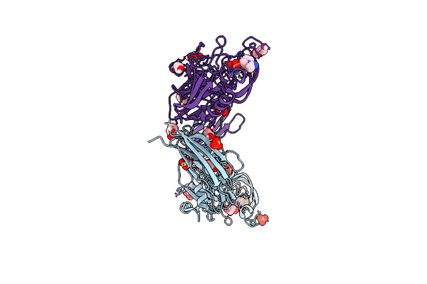
Voltage-Gated Sodium Channel 1.5 (Nav1.5) C-Terminal Domain In Complex With Calmodulin Poised For Activation
Organism: Homo sapiens
Method: X-RAY DIFFRACTION Resolution:2.80 Å Release Date: 2014-12-03 Classification: METAL BINDING PROTEIN Ligands: MG, SO4, PO4 |
 |
Crystal Structure Of M. Tuberculosis Ld-Transpeptidase Type 2 With Modified Catalytic Cysteine (C354)
Organism: Mycobacterium tuberculosis
Method: X-RAY DIFFRACTION Resolution:2.80 Å Release Date: 2012-12-12 Classification: peptidoglycan binding protein Ligands: PEG |
 |
Crystal Structure Of M. Tuberculosis Ld-Transpeptidase Type 2 Complexed With A Peptidoglycan Fragment
Organism: Mycobacterium tuberculosis
Method: X-RAY DIFFRACTION Resolution:1.72 Å Release Date: 2012-12-05 Classification: PEPTIDOGLYCAN BINDING PROTEIN Ligands: 0JC, DGL, 6CL, PT |
 |
Crystal Structure Of Mutant (C354A) M. Tuberculosis Ld-Transpeptidase Type 2
Organism: Mycobacterium tuberculosis
Method: X-RAY DIFFRACTION Resolution:2.32 Å Release Date: 2012-12-05 Classification: peptidoglycan binding protein |