
Search Count: 16
 |
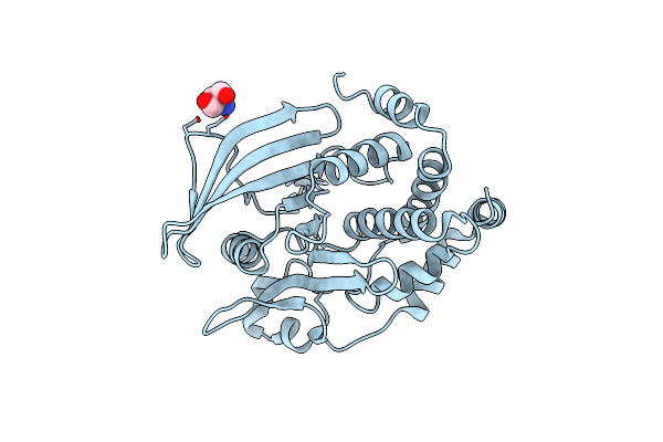
Organism: Arabidopsis thaliana
Method: X-RAY DIFFRACTION Resolution:1.30 Å Release Date: 2022-06-22 Classification: HYDROLASE |
 |
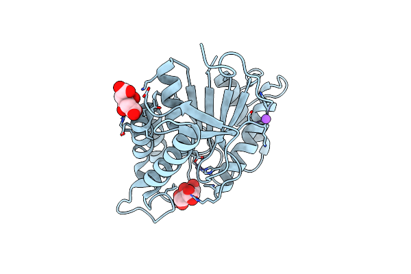
Organism: Arabidopsis thaliana
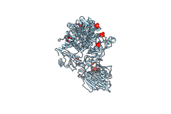
Method: X-RAY DIFFRACTION Resolution:2.03 Å Release Date: 2022-06-22 Classification: HYDROLASE Ligands: PO4 |
 |
Crystal Structure Of Full-Length Cnfy (C866S) From Yersinia Pseudotuberculosis
Organism: Yersinia pseudotuberculosis
Method: X-RAY DIFFRACTION Resolution:2.70 Å Release Date: 2020-12-30 Classification: TOXIN Ligands: SO4, CL |
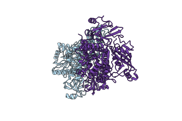 |
Crystal Structure Of Cnfy From Yersinia Pseudotuberculosis - N-Terminal Fragment Comprising Residues 1-704
Organism: Yersinia pseudotuberculosis
Method: X-RAY DIFFRACTION Resolution:3.28 Å Release Date: 2020-12-30 Classification: TOXIN |
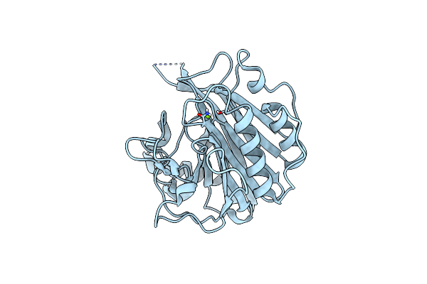 |
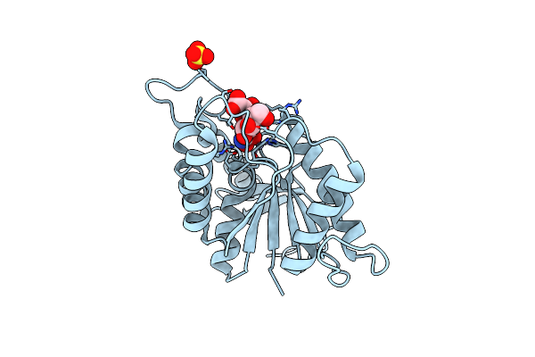
Crystal Structure Of The C-Terminal Domain Of Cnfy From Yersinia Pseudotuberculosis
Organism: Yersinia pseudotuberculosis
Method: X-RAY DIFFRACTION Resolution:1.13 Å Release Date: 2020-12-30 Classification: TOXIN Ligands: MG |
 |
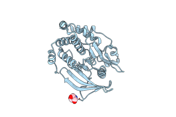
Organism: Yersinia pseudotuberculosis
Method: X-RAY DIFFRACTION Resolution:1.80 Å Release Date: 2020-12-30 Classification: TOXIN Ligands: BU3, MG, NA, CL, SO4 |
 |
Organism: Severe acute respiratory syndrome coronavirus 2, Homo sapiens
Method: ELECTRON MICROSCOPY Release Date: 2020-11-18 Classification: VIRAL PROTEIN Ligands: NAG |
 |
Organism: Bacillus subtilis (strain 168)
Method: X-RAY DIFFRACTION Resolution:1.85 Å Release Date: 2020-01-08 Classification: HYDROLASE Ligands: ZN, CIT, NA |
 |
Organism: Bacillus subtilis (strain 168)
Method: X-RAY DIFFRACTION Resolution:1.45 Å Release Date: 2020-01-08 Classification: HYDROLASE Ligands: RWI, ZN, SO4 |
 |
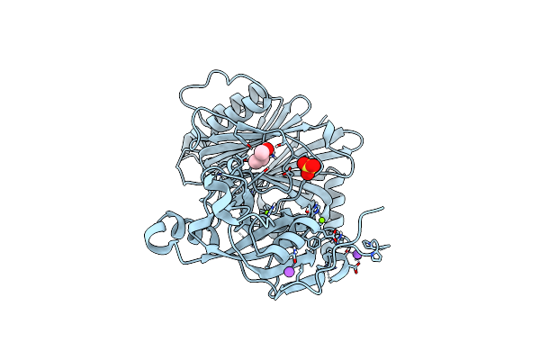
The Second Sphere Residue T263 Is Important For Function And Activity Of Ptp1B Through Modulating Wpd Loop
Organism: Homo sapiens
Method: X-RAY DIFFRACTION Resolution:1.90 Å Release Date: 2015-02-11 Classification: HYDROLASE Ligands: TRS |
 |
The Second Sphere Residue T263 Is Important For Function And Activity Of Ptp1B Through Modulating Wpd Loop
Organism: Homo sapiens
Method: X-RAY DIFFRACTION Resolution:1.91 Å Release Date: 2015-02-11 Classification: HYDROLASE Ligands: TRS |
 |
The Second Sphere Residue T263 Is Important For Function And Activity Of Ptp1B Through Modulating Wpd Loop
Organism: Homo sapiens
Method: X-RAY DIFFRACTION Resolution:2.40 Å Release Date: 2014-12-24 Classification: HYDROLASE Ligands: TRS |
 |
The Second Sphere Residue T263 Is Important For Function And Activity Of Ptp1B Through Modulating Wpd Loop
Organism: Homo sapiens
Method: X-RAY DIFFRACTION Resolution:2.29 Å Release Date: 2014-12-24 Classification: HYDROLASE |
 |
Organism: Pseudomonas aeruginosa
Method: X-RAY DIFFRACTION Resolution:1.90 Å Release Date: 2003-09-30 Classification: ELECTRON TRANSPORT Ligands: CU1, REP |
 |
Organism: Pseudomonas aeruginosa
Method: X-RAY DIFFRACTION Resolution:1.80 Å Release Date: 2001-10-17 Classification: ELECTRON TRANSPORT Ligands: CU, RTC, DPT |
 |
Organism: Gallus gallus
Method: X-RAY DIFFRACTION Resolution:1.90 Å Release Date: 1998-04-29 Classification: OXIDOREDUCTASE Ligands: SO4, MTE, MO, HEM, GOL, EPE |