
Search Count: 14
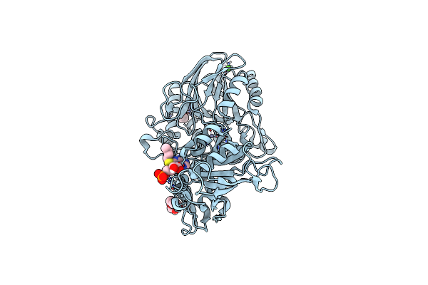 |
Crystal Structure Of Cd73 (Ecto-5'-Nucleotidase) Complexed To 8-Butylthioadenosine 5'Monophosphate (Compound 3 In Publication) In The Open Enzyme State
Organism: Homo sapiens
Method: X-RAY DIFFRACTION Resolution:1.06 Å Release Date: 2025-05-28 Classification: HYDROLASE Ligands: ZN, CA, A1IVB, 1PE, PGE |
 |
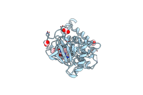
The Crystal Structure Of Inactive Magnaporthe Grisea Oxidoreductase In Complex With Nadp And Glycerol
Organism: Pyricularia grisea
Method: X-RAY DIFFRACTION Resolution:2.91 Å Release Date: 2025-03-19 Classification: OXIDOREDUCTASE Ligands: GOL, NAP |
 |
The Crystal Structure Of Wildtype Magnaporthe Grisea Oxidoreductase In Complex With Nadp
Organism: Pyricularia grisea
Method: X-RAY DIFFRACTION Resolution:2.30 Å Release Date: 2025-03-19 Classification: OXIDOREDUCTASE Ligands: NAP |
 |
Crystal Structure Of Human Tak1/Tab1 Fusion Protein In Complex With Compound S1
Organism: Homo sapiens
Method: X-RAY DIFFRACTION Resolution:2.40 Å Release Date: 2024-12-04 Classification: TRANSFERASE Ligands: A1IED, EDO |
 |
Crystal Structure Of The Activin Receptor Type-2B Ligand Binding Domain In Complex With Bimagrumab Fv, Orthorhombic Crystal Form
Organism: Homo sapiens
Method: X-RAY DIFFRACTION Resolution:2.00 Å Release Date: 2017-11-15 Classification: TRANSFERASE Ligands: PG4 |
 |
Crystal Structure Of The Activin Receptor Type-2A Ligand Binding Domain In Complex With Bimagrumab Fv
Organism: Homo sapiens
Method: X-RAY DIFFRACTION Resolution:2.35 Å Release Date: 2017-11-15 Classification: TRANSFERASE Ligands: NAG |
 |
Crystal Structure Of The Activin Receptor Type-2B Ligand Binding Domain In Complex With Bimagrumab Fv, Cubic Crystal Form
Organism: Homo sapiens
Method: X-RAY DIFFRACTION Resolution:3.35 Å Release Date: 2017-11-15 Classification: IMMUNE SYSTEM |
 |
Organism: Homo sapiens
Method: X-RAY DIFFRACTION Resolution:1.78 Å Release Date: 2017-11-15 Classification: IMMUNE SYSTEM Ligands: GOL |
 |
Organism: Vesicular stomatitis virus
Method: X-RAY DIFFRACTION Resolution:1.83 Å Release Date: 2009-01-13 Classification: VIRAL PROTEIN |
 |
Organism: Lagos bat virus
Method: X-RAY DIFFRACTION Resolution:2.75 Å Release Date: 2009-01-13 Classification: VIRAL PROTEIN |
 |
Crystal Structures Of The Small Ribosomal Subunit With Tetracycline, Edeine And If3
Organism: Thermus thermophilus
Method: X-RAY DIFFRACTION Resolution:3.20 Å Release Date: 2001-04-12 Classification: RIBOSOME Ligands: MG, WO2, ZN |
 |
Crystal Structure Of The 30S Ribosomal Subunit From Thermus Thermophilus In Complex With Edeine
Organism: Thermus thermophilus
Method: X-RAY DIFFRACTION Resolution:4.50 Å Release Date: 2001-04-12 Classification: RIBOSOME Ligands: MG, WO2, EDE, ZN |
 |
Crystal Structure Of The 30S Ribosomal Subunit From Thermus Thermophilus In Complex With The Translation Initiation Factor If3 (C-Terminal Domain)
Organism: Thermus thermophilus
Method: X-RAY DIFFRACTION Resolution:4.20 Å Release Date: 2001-04-12 Classification: RIBOSOME Ligands: MG, WO2, ZN |
 |
Crystal Structure Of The 30S Ribosomal Subunit From Thermus Thermophilus In Complex With Tetracycline
Organism: Thermus thermophilus
Method: X-RAY DIFFRACTION Resolution:4.50 Å Release Date: 2001-04-12 Classification: RIBOSOME Ligands: MG, WO2, TAC, ZN |