
Search Count: 328
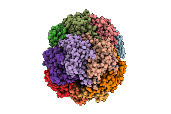 |
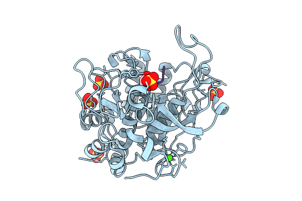
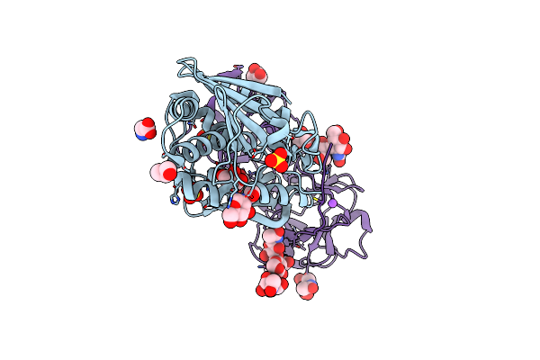
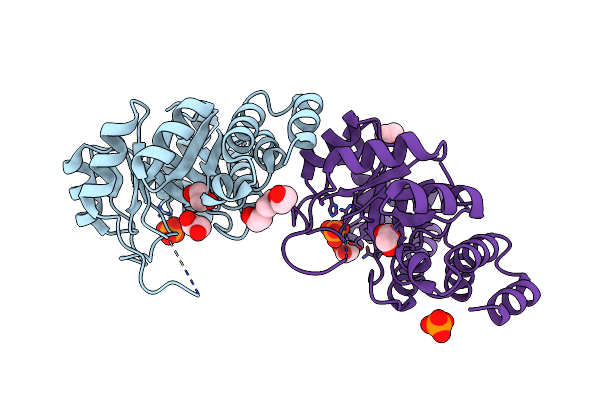
Crystal Structure Of S. Epidermidis Clpp In Complex With Tavaborole - Soaking
Organism: Staphylococcus epidermidis
Method: X-RAY DIFFRACTION Release Date: 2025-10-29 Classification: HYDROLASE Ligands: ACT, A1II0 |
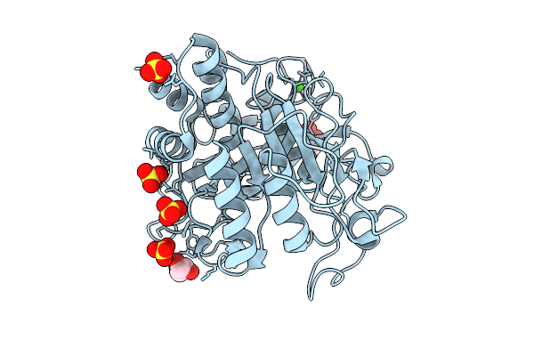 |
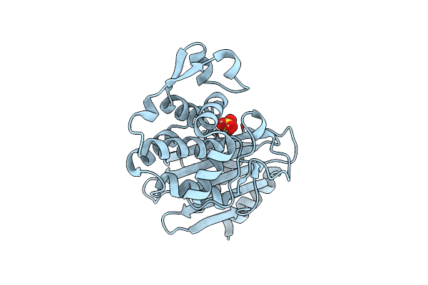
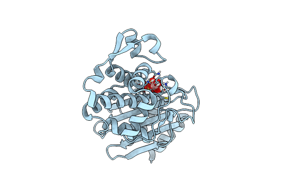
Stmpr1, Stenotrophomonas Maltophilia Protease 1, 36 Kda Alkine Serine Protease
Organism: Stenotrophomonas maltophilia
Method: X-RAY DIFFRACTION Release Date: 2025-08-06 Classification: HYDROLASE Ligands: GOL, CA, SO4 |
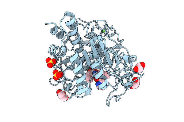 |
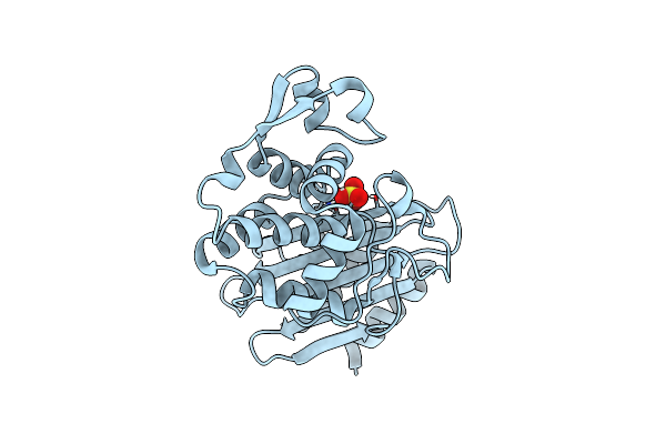
Stmpr1, Stenotrophomonas Maltophilia Protease 1, 36 Kda Alkine Serine Protease In Complex With Bortezomib
Organism: Stenotrophomonas maltophilia
Method: X-RAY DIFFRACTION Release Date: 2025-08-06 Classification: HYDROLASE Ligands: BO2, GOL, SO4, CA |
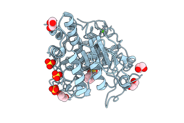 |
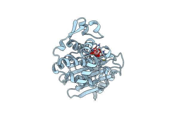
Stmpr1, Stenotrophomonas Maltophilia Protease 1, 36 Kda Alkine Serine Protease In Complex With Pmsf
Organism: Stenotrophomonas maltophilia
Method: X-RAY DIFFRACTION Release Date: 2025-08-06 Classification: HYDROLASE Ligands: GOL, PMS, SO4, CA |
 |
Stmpr1, Stenotrophomonas Maltophilia Protease 1, 36 Kda Alkine Serine Protease In Complex With Chymostatin
Organism: Stenotrophomonas maltophilia
Method: X-RAY DIFFRACTION Release Date: 2025-08-06 Classification: HYDROLASE Ligands: CA, SO4, GOL, A1I1B |
 |
Stmpr1, Stenotrophomonas Maltophilia Protease 1, 36 Kda Alkine Serine Protease In Complex With Leupeptin
Organism: Stenotrophomonas maltophilia, Synthetic construct
Method: X-RAY DIFFRACTION Release Date: 2025-08-06 Classification: HYDROLASE Ligands: CA, SO4, GOL |
 |
Crystal Structure Of S. Epidermidis Clpp In Complex With Bortezomib - Cocrystallization
Organism: Staphylococcus epidermidis
Method: X-RAY DIFFRACTION Release Date: 2025-07-30 Classification: HYDROLASE Ligands: ACT, BO2 |
 |
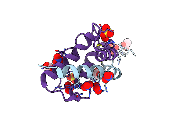
Clpp From Staphylococcus Epidermidis With Glycerol In Some Of The Catalytic Sites.
Organism: Staphylococcus epidermidis
Method: X-RAY DIFFRACTION Release Date: 2025-07-02 Classification: HYDROLASE Ligands: MPD, GOL |
 |
Crystal Structure Of Isoform Chitin Binding Protein From Iberis Umbellata L.
Organism: Iberis umbellata
Method: X-RAY DIFFRACTION Release Date: 2025-06-11 Classification: PLANT PROTEIN Ligands: SO4, NA, NO3, CL, ACT |
 |
High-Resolution Crystal Structure Of An Isoform Of Chitin Binding Protein From Iberis Umbellata L.
Organism: Iberis umbellata
Method: X-RAY DIFFRACTION Resolution:0.91 Å Release Date: 2025-03-19 Classification: PLANT PROTEIN Ligands: SO4, NO3, ACY, LI |
 |
Organism: Viscum album
Method: X-RAY DIFFRACTION Resolution:2.28 Å Release Date: 2025-01-15 Classification: PLANT PROTEIN Ligands: GOL, NAG, SO4, GLY, CL, NA |
 |
Organism: Iberis umbellata
Method: X-RAY DIFFRACTION Release Date: 2024-12-11 Classification: PLANT PROTEIN Ligands: SO4, ACT, NA, NO3, CL |
 |
Organism: Staphylococcus aureus
Method: X-RAY DIFFRACTION Resolution:2.83 Å Release Date: 2024-10-02 Classification: TRANSFERASE Ligands: PO4, EDO, CL, MG |
 |
Organism: Staphylococcus aureus
Method: X-RAY DIFFRACTION Resolution:3.02 Å Release Date: 2024-07-24 Classification: BIOSYNTHETIC PROTEIN Ligands: PO4, GLN, SO4 |
 |
Organism: Staphylococcus aureus
Method: X-RAY DIFFRACTION Resolution:3.02 Å Release Date: 2024-07-24 Classification: BIOSYNTHETIC PROTEIN Ligands: EDO, PO4, CL, PGE |
 |
Structure Of Serine-Beta-Lactamase Ctx-M-14 Following The Time-Resolved Active Site Binding Of Boric Acid, 0 Ms - Native.
Organism: Klebsiella pneumoniae
Method: X-RAY DIFFRACTION Resolution:1.40 Å Release Date: 2024-06-26 Classification: HYDROLASE Ligands: SO4 |
 |
Structure Of Serine-Beta-Lactamase Ctx-M-14 Following The Time-Resolved Active Site Binding Of Boric Acid, 50 Ms
Organism: Klebsiella pneumoniae
Method: X-RAY DIFFRACTION Resolution:1.58 Å Release Date: 2024-06-26 Classification: HYDROLASE Ligands: SO4 |
 |
Structure Of Serine-Beta-Lactamase Ctx-M-14 Following The Time-Resolved Active Site Binding Of Boric Acid, 80 Ms
Organism: Klebsiella pneumoniae
Method: X-RAY DIFFRACTION Resolution:1.69 Å Release Date: 2024-06-26 Classification: HYDROLASE Ligands: BO4, SO4 |
 |
Structure Of Serine-Beta-Lactamase Ctx-M-14 Following The Time-Resolved Active Site Binding Of Boric Acid, 100 Ms
Organism: Klebsiella pneumoniae
Method: X-RAY DIFFRACTION Resolution:2.04 Å Release Date: 2024-06-26 Classification: HYDROLASE Ligands: BO4, SO4 |
 |
Structure Of Serine-Beta-Lactamase Ctx-M-14 Following The Time-Resolved Active Site Binding Of Boric Acid, 150 Ms
Organism: Klebsiella pneumoniae
Method: X-RAY DIFFRACTION Resolution:1.97 Å Release Date: 2024-06-26 Classification: HYDROLASE Ligands: BO4, SO4 |