
Search Count: 6
All
Selected
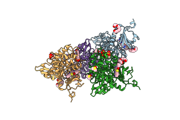 |
Organism: Homo sapiens
Method: X-RAY DIFFRACTION Resolution:2.90 Å Release Date: 2020-03-18 Classification: TRANSFERASE Ligands: SO4, PG4, DMS, JHW, EPE |
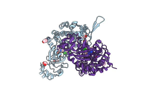 |
Organism: Homo sapiens
Method: X-RAY DIFFRACTION Resolution:2.15 Å Release Date: 2016-10-26 Classification: TRANSFERASE Ligands: 7AA, EDO |
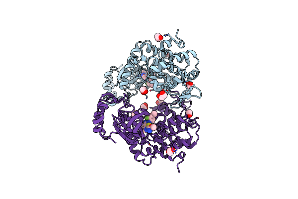 |
Organism: Homo sapiens
Method: X-RAY DIFFRACTION Resolution:2.58 Å Release Date: 2016-10-26 Classification: TRANSFERASE Ligands: 7A7, EDO |
 |
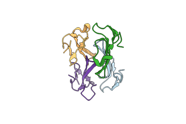
Organism: Dendroaspis polylepis polylepis
Method: X-RAY DIFFRACTION Resolution:1.70 Å Release Date: 2015-12-30 Classification: TOXIN Ligands: IOD, EDO, PGO |
 |
Crystal Structure Of Dendroaspis Polylepis Mambalgin-1 Wild-Type In P21 Space Group.
Organism: Dendroaspis polylepis polylepis
Method: X-RAY DIFFRACTION Resolution:1.80 Å Release Date: 2015-12-30 Classification: TOXIN |
 |
Crystal Structure Of Dendroaspis Polylepis Mambalgin-1 Wild-Type In P41212 Space Group
Organism: Dendroaspis polylepis polylepis
Method: X-RAY DIFFRACTION Resolution:1.95 Å Release Date: 2015-12-30 Classification: TOXIN |