
Search Count: 19
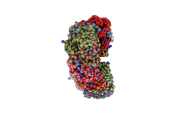 |
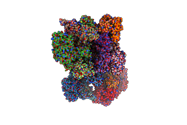
Crystal Structure Of The Membrane Domain Of Respiratory Complex I From Thermus Thermophilus
Organism: Thermus thermophilus
Method: X-RAY DIFFRACTION Resolution:3.30 Å Release Date: 2013-02-13 Classification: OXIDOREDUCTASE Ligands: UMQ |
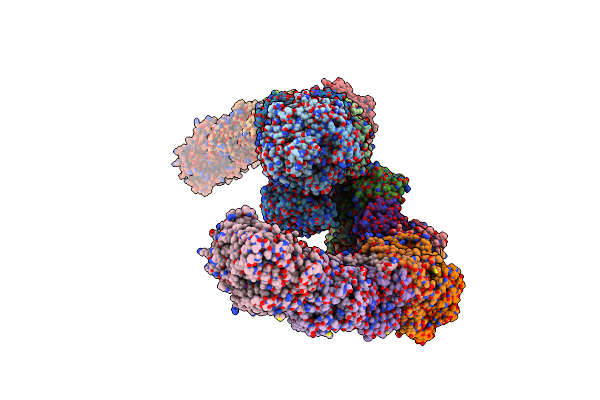 |
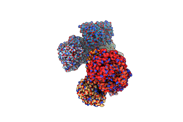
Crystal Structure Of The Entire Respiratory Complex I From Thermus Thermophilus
Organism: Thermus thermophilus
Method: X-RAY DIFFRACTION Resolution:3.30 Å Release Date: 2013-02-13 Classification: OXIDOREDUCTASE Ligands: SF4, FMN, FES |
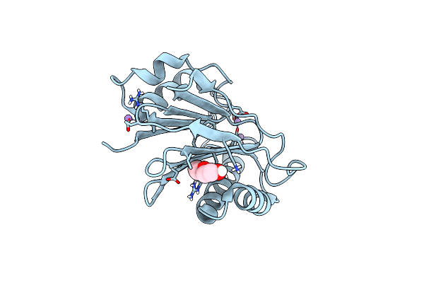 |
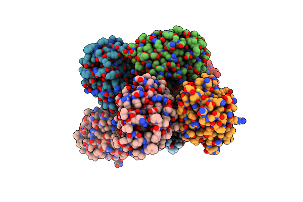
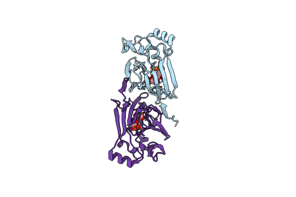
The Bacterial Stressosome: A Modular System That Has Been Adapted To Control Secondary Messenger Signaling
Organism: Moorella thermoacetica
Method: X-RAY DIFFRACTION Resolution:1.75 Å Release Date: 2012-02-22 Classification: HYDROLASE Ligands: MN, PEG |
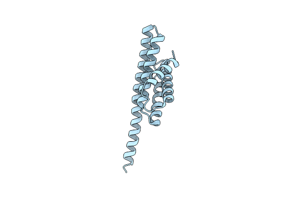 |
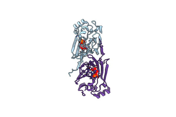
The Bacterial Stressosome: A Modular System That Has Been Adapted To Control Secondary Messenger Signaling
Organism: Moorella thermoacetica
Method: X-RAY DIFFRACTION Resolution:2.70 Å Release Date: 2012-02-22 Classification: SIGNALING |
 |
The Bacterial Stressosome: A Modular System That Has Been Adapted To Control Secondary Messenger Signaling
Organism: Moorella thermoacetica
Method: X-RAY DIFFRACTION Resolution:2.80 Å Release Date: 2012-02-22 Classification: SIGNALING Ligands: IOD |
 |
Organism: Moorella thermoacetica
Method: X-RAY DIFFRACTION Resolution:1.90 Å Release Date: 2012-02-22 Classification: TRANSCRIPTION Ligands: SCN |
 |
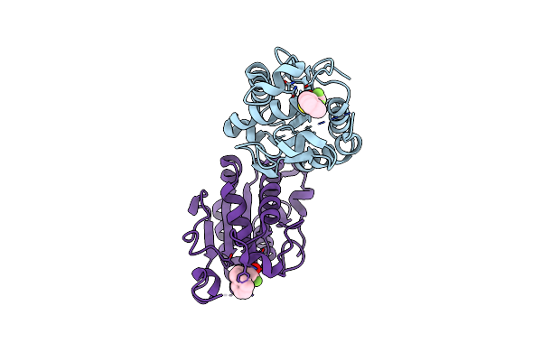
Crystal Structure Of The Hydrophilic Domain Of Respiratory Complex I From Thermus Thermophilus, Oxidized, 2 Mol/Asu
Organism: Thermus thermophilus
Method: X-RAY DIFFRACTION Resolution:3.10 Å Release Date: 2009-09-15 Classification: OXIDOREDUCTASE Ligands: SF4, FMN, FES, MN, CA |
 |
Crystal Structure Of The Hydrophilic Domain Of Respiratory Complex I From Thermus Thermophilus, Reduced, 2 Mol/Asu, With Bound Nadh
Organism: Thermus thermophilus
Method: X-RAY DIFFRACTION Resolution:3.10 Å Release Date: 2009-09-15 Classification: OXIDOREDUCTASE Ligands: SF4, FMN, NAI, MG, FES, CA |
 |
Crystal Structure Of The Hydrophilic Domain Of Respiratory Complex I From Thermus Thermophilus, Oxidized, 4 Mol/Asu, Re-Refined To 3.15 Angstrom Resolution
Organism: Thermus thermophilus
Method: X-RAY DIFFRACTION Resolution:3.15 Å Release Date: 2009-09-15 Classification: OXIDOREDUCTASE Ligands: SF4, FMN, FES, CA |
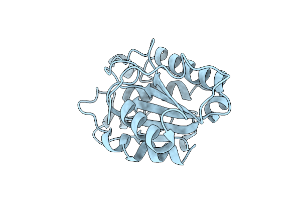 |
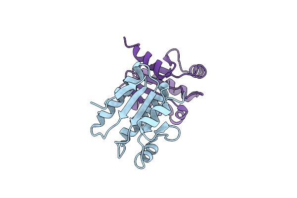
Organism: Glomerella cingulata
Method: X-RAY DIFFRACTION Resolution:1.90 Å Release Date: 2008-11-18 Classification: HYDROLASE |
 |
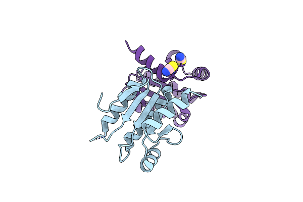
Organism: Glomerella cingulata
Method: X-RAY DIFFRACTION Resolution:2.60 Å Release Date: 2008-11-18 Classification: HYDROLASE Ligands: DEP |
 |
Organism: Glomerella cingulata
Method: X-RAY DIFFRACTION Resolution:2.30 Å Release Date: 2008-11-18 Classification: HYDROLASE Ligands: HZH |
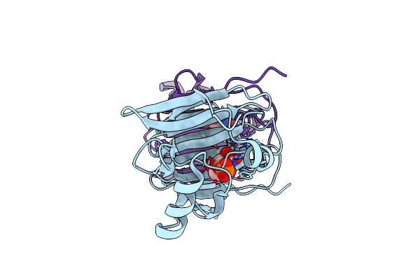 |
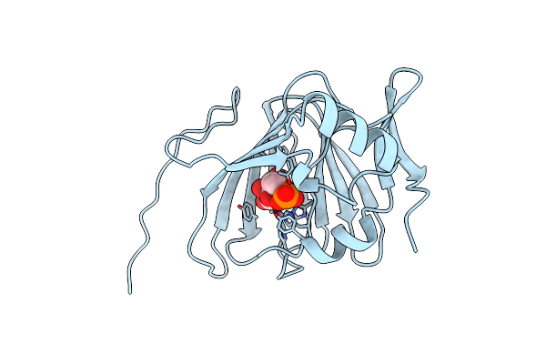
The Crystal Structure Of Phosphoglucose Isomerase From Pyrococcus Furiosus In Complex With 5-Phospho-D-Arabinonohydroxamate And Zinc
Organism: Pyrococcus furiosus
Method: X-RAY DIFFRACTION Resolution:2.00 Å Release Date: 2006-04-11 Classification: ISOMERASE Ligands: ZN, PAN |
 |
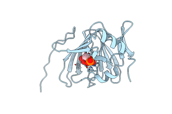
The Crystal Structure Of Phosphoglucose Isomerase From Pyrococcus Furiosus In Complex With Sorbitol 6-Phosphate And Zinc
Organism: Pyrococcus furiosus
Method: X-RAY DIFFRACTION Resolution:1.95 Å Release Date: 2006-04-11 Classification: ISOMERASE Ligands: S6P, ZN |
 |
The Crystal Structure Of Phosphoglucose Isomerase From Pyrococcus Furiosus In Complex With Fructose 6-Phosphate And Zinc
Organism: Pyrococcus furiosus
Method: X-RAY DIFFRACTION Resolution:2.10 Å Release Date: 2006-04-11 Classification: ISOMERASE Ligands: ZN, F6R |
 |
The Crystal Structure Of Phosphoglucose Isomerase From Pyrococcus Furiosus In Complex With Mannose 6-Phosphate And Zinc
Organism: Pyrococcus furiosus
Method: X-RAY DIFFRACTION Resolution:2.10 Å Release Date: 2006-04-11 Classification: ISOMERASE Ligands: ZN, M6P |
 |
The Crystal Structure Of Pyrococcus Furiosus Phosphoglucose Isomerase With Bound 5-Phospho-D-Arabinonate And Manganese
Organism: Pyrococcus furiosus
Method: X-RAY DIFFRACTION Resolution:1.89 Å Release Date: 2004-10-12 Classification: ISOMERASE Ligands: MN, PA5 |
 |
Crystal Structure Of Phosphoglucose Isomerase From Pyrococcus Furiosus With Bound 5-Phospho-D-Arabinonate
Organism: Pyrococcus furiosus
Method: X-RAY DIFFRACTION Resolution:1.50 Å Release Date: 2004-10-12 Classification: METAL BINDING PROTEIN Ligands: PA5 |
 |
Crystal Structure Of Pyrococcus Furiosus Phosphoglucose Isomerase Free Enzyme
Organism: Pyrococcus furiosus
Method: X-RAY DIFFRACTION Resolution:2.80 Å Release Date: 2004-10-12 Classification: ISOMERASE |