
Search Count: 35
 |
Organism: Homo sapiens
Method: X-RAY DIFFRACTION Release Date: 2025-07-30 Classification: SUGAR BINDING PROTEIN Ligands: A1IIW, CA, CL, TRS |
 |
Organism: Pseudomonas aeruginosa pao1
Method: X-RAY DIFFRACTION Resolution:1.55 Å Release Date: 2025-04-16 Classification: SUGAR BINDING PROTEIN Ligands: CA, SO4, R7E |
 |
Organism: Pseudomonas aeruginosa pao1
Method: X-RAY DIFFRACTION Resolution:1.74 Å Release Date: 2025-04-16 Classification: SUGAR BINDING PROTEIN Ligands: CA, R7E, A1IH4 |
 |
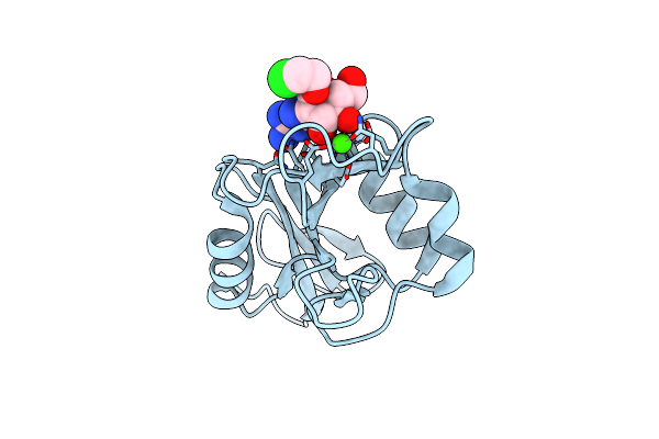
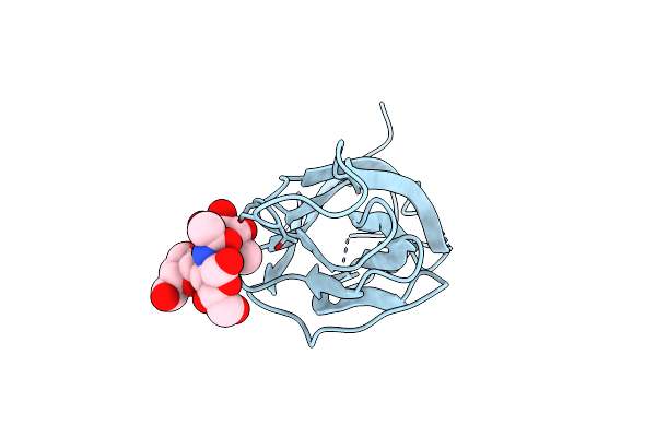
N Terminal Domain Of Bc2L-C Lectin In Complex With N-(Beta-L-Fucopyranosyl)-Biphenyl-3-Carboxamide
Organism: Burkholderia cenocepacia j2315
Method: X-RAY DIFFRACTION Resolution:2.55 Å Release Date: 2025-04-16 Classification: SUGAR BINDING PROTEIN Ligands: MJO, PG4, 1PE |
 |
Organism: Homo sapiens
Method: X-RAY DIFFRACTION Resolution:1.80 Å Release Date: 2024-10-16 Classification: SUGAR BINDING PROTEIN Ligands: CA, CL, A1H0Q |
 |
Structure Of The N-Terminal Domain Of Bc2L-C Lectin (1-131) In Complex With A Synthetic Beta-Fucosylamide
Organism: Burkholderia cenocepacia
Method: X-RAY DIFFRACTION Resolution:1.55 Å Release Date: 2023-02-08 Classification: SUGAR BINDING PROTEIN Ligands: R7E |
 |
Structure Of The N-Terminal Domain Of Bc2L-C Lectin (1-131) In Complex With A Synthetic Beta-C-Fucoside Ligand
Organism: Burkholderia cenocepacia (strain atcc baa-245 / dsm 16553 / lmg 16656 / nctc 13227 / j2315 / cf5610)
Method: X-RAY DIFFRACTION Resolution:1.79 Å Release Date: 2022-06-01 Classification: SUGAR BINDING PROTEIN Ligands: VJT |
 |
Structure Of The N-Terminal Domain Of Bc2L-C Lectin (1-131) In Complex With A Synthetic Beta-N-Fucoside Ligand
Organism: Burkholderia cenocepacia (strain atcc baa-245 / dsm 16553 / lmg 16656 / nctc 13227 / j2315 / cf5610)
Method: X-RAY DIFFRACTION Resolution:1.32 Å Release Date: 2022-06-01 Classification: SUGAR BINDING PROTEIN Ligands: VJW |
 |
Structure Of The N Terminal Domain Of Bc2L-C Lectin (1-131) In Complex With Globo H (H-Type 3) And Cas No 912569-62-1
Organism: Burkholderia cenocepacia (strain atcc baa-245 / dsm 16553 / lmg 16656 / nctc 13227 / j2315 / cf5610)
Method: X-RAY DIFFRACTION Resolution:1.90 Å Release Date: 2021-04-07 Classification: SUGAR BINDING PROTEIN Ligands: QT5, NA |
 |
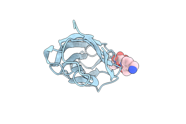
Structure Of The Apo Form Of The N Terminal Domain Of Bc2L-C Lectin (1-131)
Organism: Burkholderia cenocepacia j2315
Method: X-RAY DIFFRACTION Resolution:1.50 Å Release Date: 2021-04-07 Classification: SUGAR BINDING PROTEIN |
 |
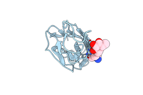
Structure Of The N Terminal Domain Of Bc2L-C Lectin (1-131) In Complex With H-Type 1 Antigen
Organism: Burkholderia cenocepacia (strain atcc baa-245 / dsm 16553 / lmg 16656 / nctc 13227 / j2315 / cf5610)
Method: X-RAY DIFFRACTION Resolution:1.61 Å Release Date: 2020-01-22 Classification: SUGAR BINDING PROTEIN |
 |
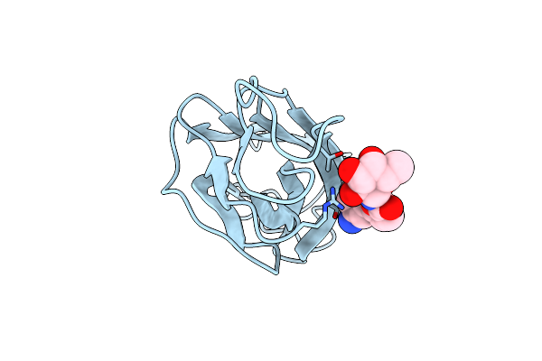
Structure Of The N Terminal Domain Of Bc2L-C Lectin (1-131) In Complex With Globo H (H-Type 3) Antigen
Organism: Burkholderia cenocepacia j2315
Method: X-RAY DIFFRACTION Resolution:1.90 Å Release Date: 2020-01-22 Classification: SUGAR BINDING PROTEIN |
 |
Organism: Homo sapiens
Method: X-RAY DIFFRACTION Resolution:2.10 Å Release Date: 2019-09-11 Classification: SUGAR BINDING PROTEIN Ligands: EZ8, CA, CL |
 |
Organism: Homo sapiens
Method: X-RAY DIFFRACTION Resolution:1.84 Å Release Date: 2018-02-21 Classification: CARBOHYDRATE BINDING PROTEIN Ligands: TRP, SGN, EU, CA, CL |
 |
Organism: Vibrio cholerae serotype o1 (strain atcc 39315 / el tor inaba n16961)
Method: X-RAY DIFFRACTION Resolution:1.13 Å Release Date: 2017-05-31 Classification: TOXIN Ligands: 7BN, TRS, IMD, PEG, MRD |
 |
Organism: Vibrio cholerae
Method: X-RAY DIFFRACTION Resolution:1.13 Å Release Date: 2017-05-31 Classification: TOXIN Ligands: 7BN, MRD, CA, TRS, PEG |
 |
Organism: Escherichia coli
Method: X-RAY DIFFRACTION Resolution:1.60 Å Release Date: 2017-05-31 Classification: TOXIN Ligands: GOL, 7BQ, PEG, 7DB, PO4 |
 |
Organism: Vibrio cholerae serotype o1 (strain atcc 39315 / el tor inaba n16961)
Method: X-RAY DIFFRACTION Resolution:1.20 Å Release Date: 2017-05-31 Classification: TOXIN Ligands: SO4, PEG, PGE, 7BT |
 |
Crystal Structure Of Adenovirus 8 Protease In Complex With A Nitrile Inhibitor
Organism: Synthetic construct, Human adenovirus d serotype 8
Method: X-RAY DIFFRACTION Resolution:1.03 Å Release Date: 2015-01-14 Classification: HYDROLASE Ligands: 3VF, EPE, GLY |
 |
Crystal Structure Of Human Adenovirus 8 Protease With An Irreversible Vinyl Sulfone Inhibitor
Organism: Human adenovirus d serotype 8
Method: X-RAY DIFFRACTION Resolution:2.15 Å Release Date: 2015-01-14 Classification: HYDROLASE Ligands: 3VK |