Search Count: 59
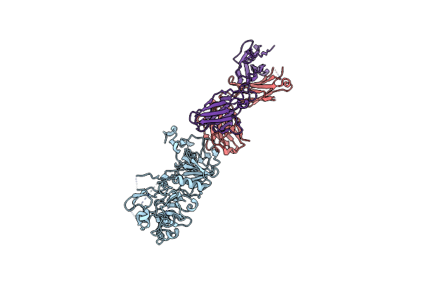 |
Organism: Plasmodium vivax sal-1, Mus musculus
Method: X-RAY DIFFRACTION Resolution:3.05 Å Release Date: 2024-10-16 Classification: IMMUNE SYSTEM |
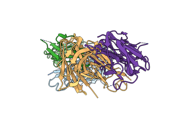 |
Organism: Mus musculus
Method: X-RAY DIFFRACTION Resolution:2.10 Å Release Date: 2024-10-16 Classification: IMMUNE SYSTEM Ligands: CL |
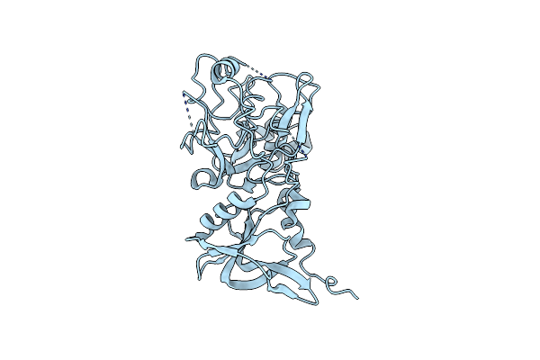 |
Organism: Plasmodium falciparum
Method: X-RAY DIFFRACTION Resolution:2.00 Å Release Date: 2024-10-16 Classification: CELL INVASION Ligands: CL |
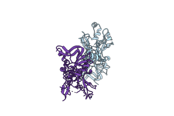 |
Organism: Plasmodium falciparum 3d7
Method: X-RAY DIFFRACTION Resolution:2.10 Å Release Date: 2024-10-16 Classification: CELL INVASION |
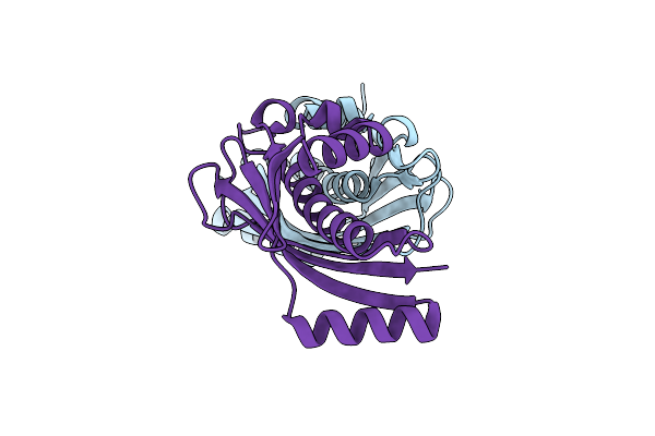 |
Crystal Structure Of The Malonyl-Acp Decarboxylase Madb From Pseudomonas Putida
Organism: Pseudomonas putida kt2440
Method: X-RAY DIFFRACTION Resolution:1.04 Å Release Date: 2023-03-01 Classification: TRANSFERASE |
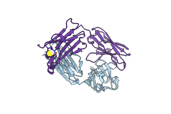 |
Organism: Mus musculus
Method: X-RAY DIFFRACTION Resolution:1.89 Å Release Date: 2021-04-14 Classification: IMMUNE SYSTEM Ligands: ZN, SCN |
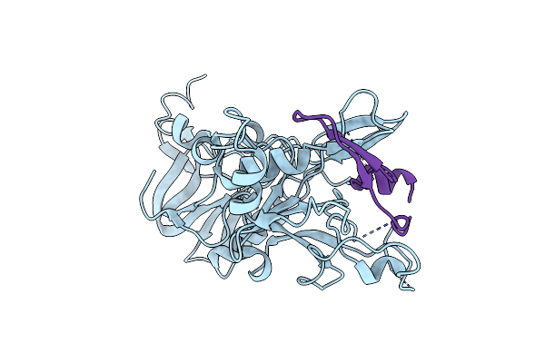 |
Crystal Structure Of Plasmodium Falciparum Ama1 In Complex With A 39 Aa Pvron2 Peptide
Organism: Plasmodium falciparum vietnam oak-knoll (fvo), Plasmodium vivax sal-1
Method: X-RAY DIFFRACTION Resolution:1.90 Å Release Date: 2017-09-06 Classification: CELL INVASION |
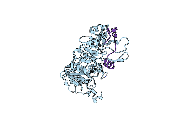 |
Crystal Structure Of Plasmodium Vivax Ama1 In Complex With A 39 Aa Pvron2 Peptide
Organism: Plasmodium vivax sal-1, Plasmodium vivax
Method: X-RAY DIFFRACTION Resolution:2.15 Å Release Date: 2017-09-06 Classification: CELL INVASION |
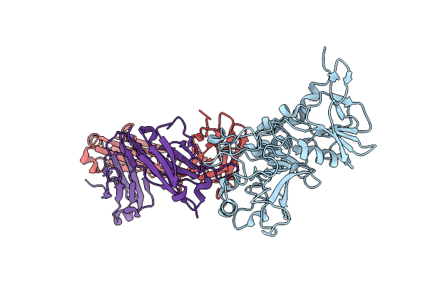 |
Crystal Structure Of Apical Membrane Antigen 1 From Plasmodium Knowlesi In Complex With An Invasion Inhibitory Antibody
Organism: Plasmodium knowlesi, Rattus norvegicus
Method: X-RAY DIFFRACTION Resolution:3.10 Å Release Date: 2015-04-29 Classification: CELL INVASION |
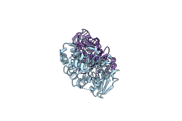 |
Organism: Plasmodium knowlesi
Method: X-RAY DIFFRACTION Resolution:2.45 Å Release Date: 2015-04-29 Classification: CELL INVASION |
 |
Crystal Structure Of The Dbl3X-Dbl4Epsilon Double Domain From The Extracellular Part Of Var2Csa Pfemp1 From Plasmodium Falciparum
Organism: Plasmodium falciparum
Method: X-RAY DIFFRACTION Resolution:2.90 Å Release Date: 2015-03-04 Classification: MEMBRANE PROTEIN |
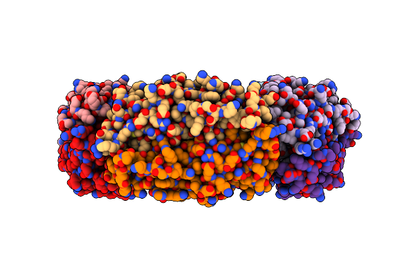 |
Tryparedoxin Peroxidase (Txnpx) From Trypanosoma Cruzi In The Reduced State
Organism: Trypanosoma cruzi
Method: X-RAY DIFFRACTION Resolution:2.80 Å Release Date: 2013-10-09 Classification: OXIDOREDUCTASE |
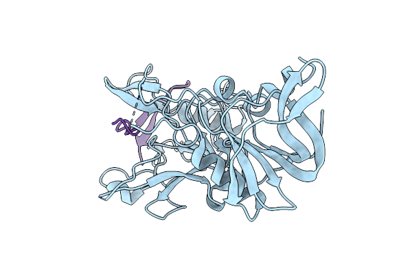 |
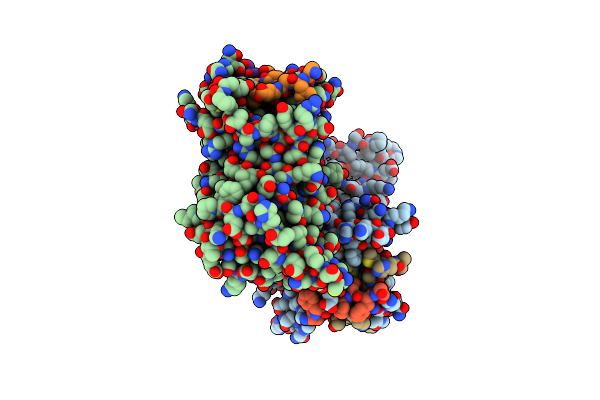
Crystal Structure Of Plasmodium Falciparum Ama1 In Complex With A 29Aa Pfron2 Peptide
Organism: Plasmodium falciparum
Method: X-RAY DIFFRACTION Resolution:1.60 Å Release Date: 2012-07-11 Classification: CELL INVASION |
 |
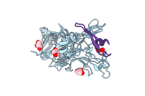
Organism: Plasmodium falciparum
Method: X-RAY DIFFRACTION Resolution:2.15 Å Release Date: 2012-07-11 Classification: CELL INVASION/inhibitor |
 |
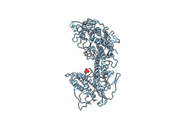
Crystal Structure Of Plasmodium Falciparum Ama1 In Complex With A 39Aa Pfron2 Peptide
Organism: Plasmodium falciparum
Method: X-RAY DIFFRACTION Resolution:2.10 Å Release Date: 2012-07-11 Classification: IMMUNE SYSTEM Ligands: GOL |
 |
Structure Of The N-Terminal Nts-Dbl1-Alpha And Cidr-Gamma Double Domain Of The Pfemp1 Protein From Plasmodium Falciparum Varo Strain.
Organism: Plasmodium falciparum
Method: X-RAY DIFFRACTION Resolution:2.80 Å Release Date: 2012-05-30 Classification: MEMBRANE PROTEIN Ligands: MG, SO4 |
 |
Organism: Plasmodium falciparum
Method: X-RAY DIFFRACTION Resolution:1.84 Å Release Date: 2012-02-15 Classification: MEMBRANE PROTEIN Ligands: DHL, BEN |
 |
Organism: Mus musculus
Method: X-RAY DIFFRACTION Resolution:2.31 Å Release Date: 2011-11-09 Classification: IMMUNE SYSTEM Ligands: CA |
 |
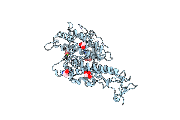
Organism: Mus musculus
Method: X-RAY DIFFRACTION Resolution:1.89 Å Release Date: 2011-11-09 Classification: IMMUNE SYSTEM Ligands: CA |
 |
Crystal Structure Of The Nts-Dbl1(Alpha-1) Domain Of The Plasmodium Falciparum Membrane Protein 1 (Pfemp1) From The Varo Strain.
Organism: Plasmodium falciparum palo alto/uganda
Method: X-RAY DIFFRACTION Resolution:2.06 Å Release Date: 2011-04-06 Classification: MEMBRANE PROTEIN Ligands: PRO, MG, GOL |

