
Search Count: 22
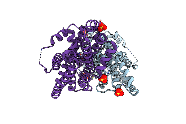 |
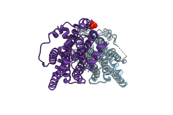
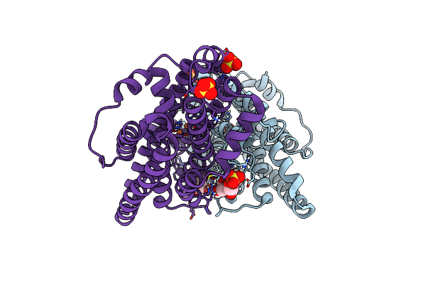
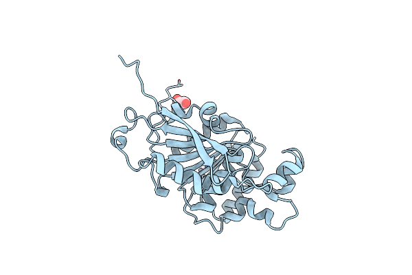
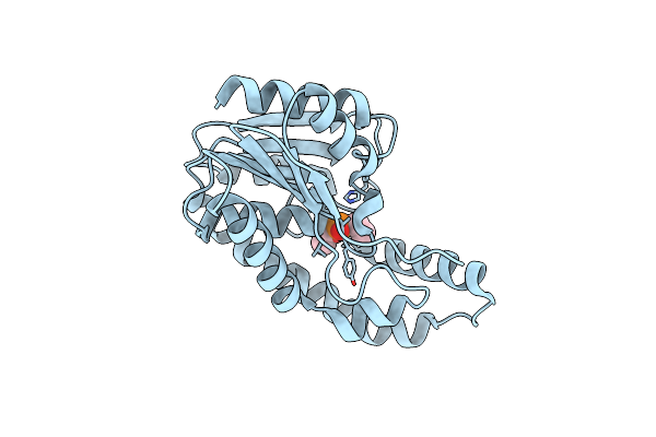
Crystal Structure Of Ribonucleotide Reductase Nrdf From Bacillus Anthracis Aerobically Soaked With Fe(Ii) And Mn(Ii) Ions
Organism: Bacillus anthracis str. sterne
Method: X-RAY DIFFRACTION Resolution:1.35 Å Release Date: 2020-04-15 Classification: OXIDOREDUCTASE Ligands: SO4, MN |
 |
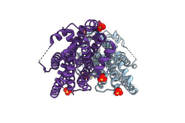
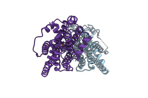
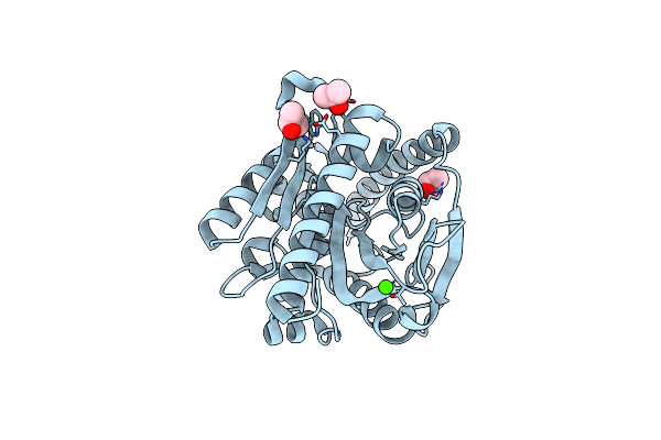
Crystal Structure Of Ribonucleotide Reductase Nrdf From Bacillus Anthracis Anaerobically Soaked With Fe(Ii) And Mn(Ii) Ions
Organism: Bacillus anthracis str. sterne
Method: X-RAY DIFFRACTION Resolution:1.55 Å Release Date: 2020-04-15 Classification: OXIDOREDUCTASE Ligands: MN, SO4 |
 |
Crystal Structure Of Apo (Metal-Free) Ribonucleotide Reductase Nrdf L61G Variant From Bacillus Anthracis
Organism: Bacillus anthracis
Method: X-RAY DIFFRACTION Resolution:2.05 Å Release Date: 2020-04-15 Classification: OXIDOREDUCTASE Ligands: SO4 |
 |
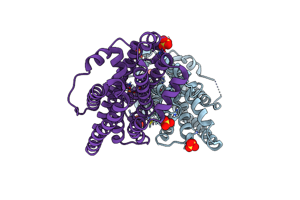
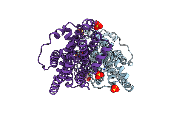
Crystal Structure Of Ribonucleotide Reductase Nrdf L61G Variant From Bacillus Anthracis Anaerobically Soaked With Fe(Ii) And Mn(Ii) Ions
Organism: Bacillus anthracis
Method: X-RAY DIFFRACTION Resolution:1.77 Å Release Date: 2020-04-15 Classification: OXIDOREDUCTASE Ligands: MN, SO4 |
 |
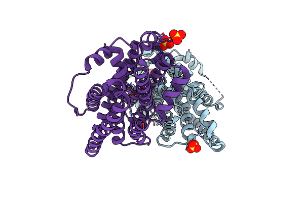
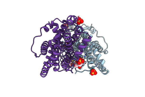
Crystal Structure Of Ribonucleotide Reductase Nrdf L61G Variant From Bacillus Anthracis Aerobically Soaked With Fe(Ii) And Mn(Ii) Ions
Organism: Bacillus anthracis
Method: X-RAY DIFFRACTION Resolution:2.10 Å Release Date: 2020-04-15 Classification: OXIDOREDUCTASE Ligands: FE2, SO4, MN |
 |
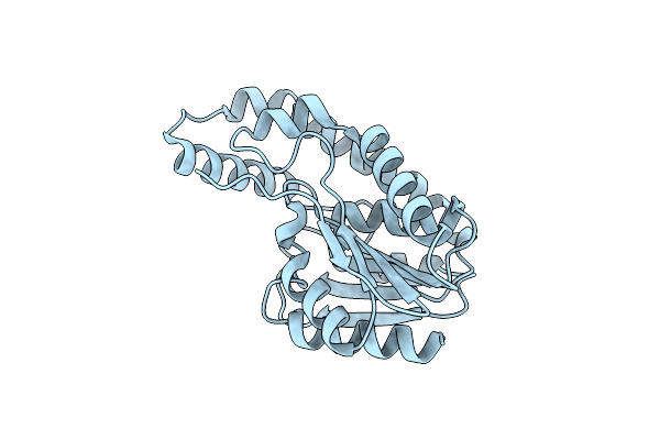
Crystal Structure Of Apo (Metal-Free) Ribonucleotide Reductase Nrdf From Bacillus Anthracis
Organism: Bacillus anthracis
Method: X-RAY DIFFRACTION Resolution:1.51 Å Release Date: 2019-08-21 Classification: OXIDOREDUCTASE Ligands: CL, SO4 |
 |
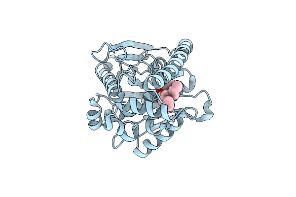
Crystal Structure Of Ribonucleotide Reductase Nrdf From Bacillus Anthracis Aerobically Soaked With Ferrous Ions (Photo-Reduced)
Organism: Bacillus anthracis str. sterne
Method: X-RAY DIFFRACTION Resolution:1.63 Å Release Date: 2019-08-21 Classification: OXIDOREDUCTASE Ligands: FE2, CL |
 |
Crystal Structure Of Ribonucleotide Reductase Nrdf From Bacillus Anthracis Anaerobically Soaked With Ferrous Ions
Organism: Bacillus anthracis str. sterne
Method: X-RAY DIFFRACTION Resolution:1.32 Å Release Date: 2019-08-21 Classification: OXIDOREDUCTASE Ligands: FE2, SO4, BTB |
 |
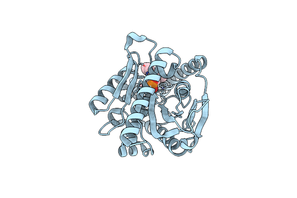
Crystal Structure Of Ribonucleotide Reductase Nrdf From Bacillus Anthracis Soaked With Manganese Ions
Organism: Bacillus anthracis
Method: X-RAY DIFFRACTION Resolution:1.30 Å Release Date: 2019-08-21 Classification: OXIDOREDUCTASE Ligands: SO4, BTB, MN |
 |
Crystal Structure Of Ribonucleotide Reductase Nrdf From Bacillus Anthracis With Partially Oxidised Di-Iron Metallocofactor
Organism: Bacillus anthracis
Method: X-RAY DIFFRACTION Resolution:1.46 Å Release Date: 2019-08-21 Classification: OXIDOREDUCTASE Ligands: SO4, FE, BTB |
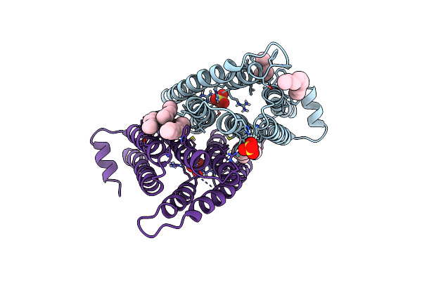 |
Crystal Structure Of Mycobacterium Tuberculosis Phosphatidylinositol Phosphate Synthase (Pgsa1) In Apo Form
Organism: mycobacterium tuberculosis h37rv
Method: X-RAY DIFFRACTION Resolution:2.90 Å Release Date: 2019-05-15 Classification: TRANSFERASE Ligands: SO4, UPL |
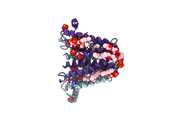 |
Crystal Structure Of Mycobacterium Tuberculosis Phosphatidylinositol Phosphate Synthase (Pgsa1) With Cdp-Dag Bound
Organism: Mycobacterium tuberculosis (strain atcc 25618 / h37rv)
Method: X-RAY DIFFRACTION Resolution:1.80 Å Release Date: 2019-05-15 Classification: TRANSFERASE Ligands: MG, SO4, PGE, FQT, OLB, 1PE |
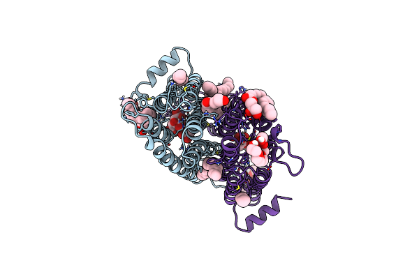 |
Crystal Structure Of Mycobacterium Tuberculosis Phosphatidylinositol Phosphate Synthase (Pgsa1) In Complex With Manganese And Citrate
Organism: Mycobacterium tuberculosis (strain atcc 25618 / h37rv)
Method: X-RAY DIFFRACTION Resolution:1.88 Å Release Date: 2019-05-15 Classification: TRANSFERASE Ligands: MN, FLC, OLC, LFA, SO4, 1PE |
 |
Organism: Streptococcus pyogenes
Method: X-RAY DIFFRACTION Resolution:1.80 Å Release Date: 2014-12-24 Classification: HYDROLASE Ligands: ZN, OXL |
 |
Organism: Lactobacillus rhamnosus
Method: X-RAY DIFFRACTION Resolution:1.71 Å Release Date: 2014-10-15 Classification: HYDROLASE Ligands: IPA, CL, CA |
 |
Crystal Structure Of A C6-C4 Sn3 Inhibited Esterase B From Lactobacillus Rhamnosis
Organism: Lactobacillus rhamnosus
Method: X-RAY DIFFRACTION Resolution:2.00 Å Release Date: 2014-08-27 Classification: HYDROLASE Ligands: C63 |
 |
Crystal Structure Of A C4-C4 Sn3 Tributyrin Phosphonate Inhibited By Esterase B From Lactobacillus Rhamnosis
Organism: Lactobacillus rhamnosus
Method: X-RAY DIFFRACTION Resolution:1.96 Å Release Date: 2014-06-11 Classification: HYDROLASE Ligands: HY4 |
 |
Snapshots Of Esterase D From Lactobacillus Rhamnosus: Insights Into A Rotation Driven Catalytic Mechanism
Organism: Lactobacillus rhamnosus
Method: X-RAY DIFFRACTION Resolution:2.20 Å Release Date: 2009-08-04 Classification: HYDROLASE Ligands: DEP |
 |
Snapshots Of Esterase D From Lactobacillus Rhamnosus: Insights Into A Rotation Driven Catalytic Mechanism
Organism: Lactobacillus rhamnosus
Method: X-RAY DIFFRACTION Resolution:1.72 Å Release Date: 2009-07-28 Classification: HYDROLASE Ligands: CL |
 |
Snapshots Of Esterase D From Lactobacillus Rhamnosus: Insights Into A Rotation Driven Catalytic Mechanism
Organism: Lactobacillus rhamnosus
Method: X-RAY DIFFRACTION Resolution:1.81 Å Release Date: 2009-07-28 Classification: HYDROLASE Ligands: CL |