
Search Count: 12
 |
Organism: Homo sapiens
Method: X-RAY DIFFRACTION Release Date: 2025-07-30 Classification: SUGAR BINDING PROTEIN Ligands: A1IIW, CA, CL, TRS |
 |
Organism: Pseudomonas aeruginosa pao1
Method: X-RAY DIFFRACTION Resolution:1.55 Å Release Date: 2025-04-16 Classification: SUGAR BINDING PROTEIN Ligands: CA, SO4, R7E |
 |
Organism: Pseudomonas aeruginosa pao1
Method: X-RAY DIFFRACTION Resolution:1.74 Å Release Date: 2025-04-16 Classification: SUGAR BINDING PROTEIN Ligands: CA, R7E, A1IH4 |
 |
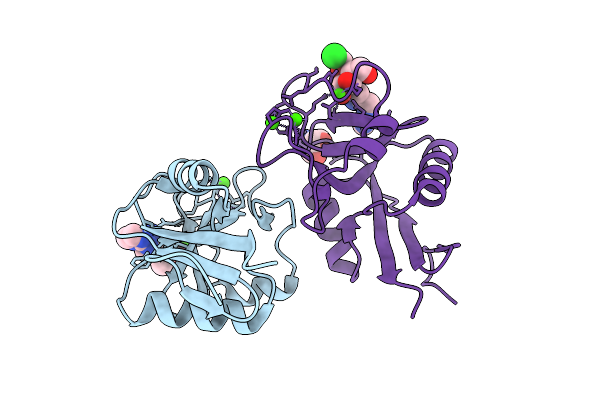
N Terminal Domain Of Bc2L-C Lectin In Complex With N-(Beta-L-Fucopyranosyl)-Biphenyl-3-Carboxamide
Organism: Burkholderia cenocepacia j2315
Method: X-RAY DIFFRACTION Resolution:2.55 Å Release Date: 2025-04-16 Classification: SUGAR BINDING PROTEIN Ligands: MJO, PG4, 1PE |
 |
Organism: Homo sapiens
Method: X-RAY DIFFRACTION Resolution:1.80 Å Release Date: 2024-10-16 Classification: SUGAR BINDING PROTEIN Ligands: CA, CL, A1H0Q |
 |
Structure Of The N-Terminal Domain Of Bc2L-C Lectin (1-131) In Complex With A Synthetic Beta-Fucosylamide
Organism: Burkholderia cenocepacia
Method: X-RAY DIFFRACTION Resolution:1.55 Å Release Date: 2023-02-08 Classification: SUGAR BINDING PROTEIN Ligands: R7E |
 |
Structure Of The N-Terminal Domain Of Bc2L-C Lectin (1-131) In Complex With A Synthetic Beta-C-Fucoside Ligand
Organism: Burkholderia cenocepacia (strain atcc baa-245 / dsm 16553 / lmg 16656 / nctc 13227 / j2315 / cf5610)
Method: X-RAY DIFFRACTION Resolution:1.79 Å Release Date: 2022-06-01 Classification: SUGAR BINDING PROTEIN Ligands: VJT |
 |
Structure Of The N-Terminal Domain Of Bc2L-C Lectin (1-131) In Complex With A Synthetic Beta-N-Fucoside Ligand
Organism: Burkholderia cenocepacia (strain atcc baa-245 / dsm 16553 / lmg 16656 / nctc 13227 / j2315 / cf5610)
Method: X-RAY DIFFRACTION Resolution:1.32 Å Release Date: 2022-06-01 Classification: SUGAR BINDING PROTEIN Ligands: VJW |
 |
Structure Of The N Terminal Domain Of Bc2L-C Lectin (1-131) In Complex With Globo H (H-Type 3) And Cas No 912569-62-1
Organism: Burkholderia cenocepacia (strain atcc baa-245 / dsm 16553 / lmg 16656 / nctc 13227 / j2315 / cf5610)
Method: X-RAY DIFFRACTION Resolution:1.90 Å Release Date: 2021-04-07 Classification: SUGAR BINDING PROTEIN Ligands: QT5, NA |
 |
Structure Of The Apo Form Of The N Terminal Domain Of Bc2L-C Lectin (1-131)
Organism: Burkholderia cenocepacia j2315
Method: X-RAY DIFFRACTION Resolution:1.50 Å Release Date: 2021-04-07 Classification: SUGAR BINDING PROTEIN |
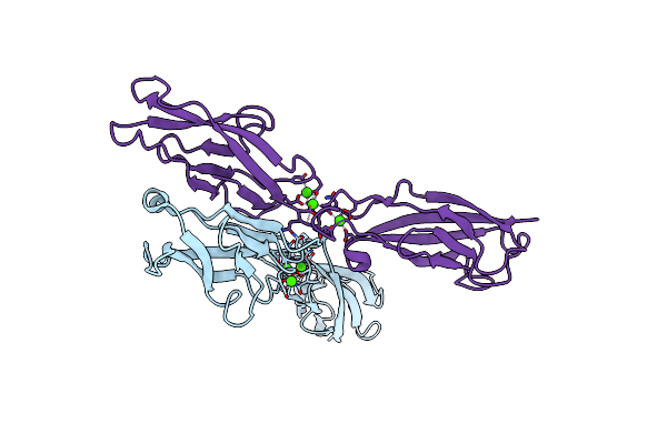 |
Crystal Structure Of Human E-Cadherin (Residues 3-213) In X-Dimer Conformation
Organism: Homo sapiens
Method: X-RAY DIFFRACTION Resolution:1.92 Å Release Date: 2016-06-01 Classification: CELL ADHESION Ligands: CA |
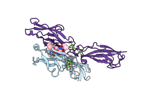 |
Crystal Structure Of Human E-Cadherin (Residues 3-213) In Complex With A Peptidomimetic Inhibitor
Organism: Homo sapiens
Method: X-RAY DIFFRACTION Resolution:2.13 Å Release Date: 2016-06-01 Classification: CELL ADHESION Ligands: CA, 4RL |