
Search Count: 88
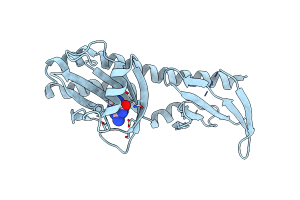 |
Crystal Structure Of The Ligand Binding Domain Of The Halomonas Titanicae Chemoreceptor Htc10 In Complex With Guanine
Organism: Halomonas titanicae
Method: X-RAY DIFFRACTION Resolution:3.60 Å Release Date: 2024-11-20 Classification: SIGNALING PROTEIN Ligands: GUN |
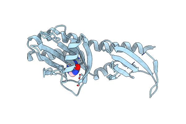 |
Crystal Structure Of The Ligand Binding Domain Of The Halomonas Titanicae Chemoreceptor Htc10 In Complex With Hypoxanthine
Organism: Halomonas titanicae
Method: X-RAY DIFFRACTION Resolution:3.63 Å Release Date: 2024-11-20 Classification: SIGNALING PROTEIN Ligands: HPA |
 |
High-Resolution Crystal Structure Of The Ligand Binding Domain Of The Halomonas Titanicae Chemoreceptor Htc10 In Complex With Guanine
Organism: Halomonas titanicae
Method: X-RAY DIFFRACTION Resolution:2.10 Å Release Date: 2024-11-20 Classification: SIGNALING PROTEIN Ligands: GUN |
 |
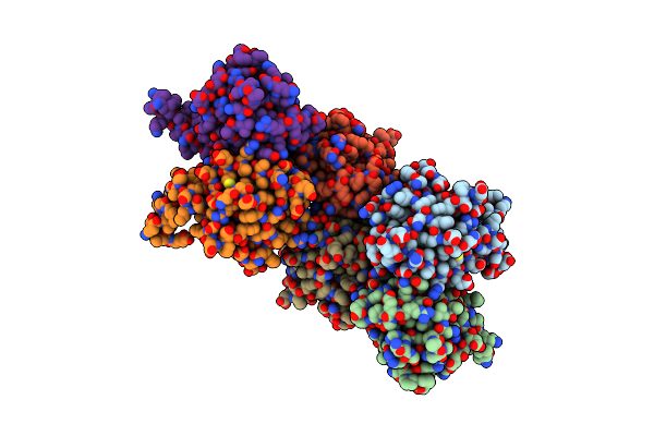
Corynebacterium Glutamicum Pyruvate:Quinone Oxidoreductase (Pqo) Purified From Bacteria Grown In Acetate Minimal Medium
Organism: Corynebacterium glutamicum atcc 13032
Method: X-RAY DIFFRACTION Resolution:1.22 Å Release Date: 2024-08-07 Classification: OXIDOREDUCTASE Ligands: FAD, TPP, MG |
 |
Pyruvate:Quinone Oxidoreductase (Pqo) From Corynebacterium Glutamicum Cs176
Organism: Corynebacterium glutamicum
Method: X-RAY DIFFRACTION Resolution:1.47 Å Release Date: 2024-08-07 Classification: OXIDOREDUCTASE Ligands: FAD |
 |
Corynebacterium Glutamicum Cs176 Pyruvate:Quinone Oxidoreductase (Pqo) In Complex With Fad And Thiamine Diphosphate-Magnesium Ion
Organism: Corynebacterium glutamicum
Method: X-RAY DIFFRACTION Resolution:1.86 Å Release Date: 2024-08-07 Classification: OXIDOREDUCTASE Ligands: TPP, MG, FAD |
 |
Corynebacterium Glutamicum Pyruvate:Quinone Oxidoreductase (Pqo), C-Terminal Truncated Construct
Organism: Corynebacterium glutamicum atcc 13032
Method: X-RAY DIFFRACTION Resolution:1.89 Å Release Date: 2024-08-07 Classification: OXIDOREDUCTASE Ligands: FAD, TPP, MG |
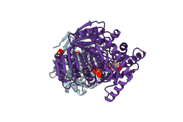 |
M. Tuberculosis Salicylate Synthase Mbti In Complex With 5-(3-Carboxyphenyl)Furan-2-Carboxylic Acid
Organism: Mycobacterium tuberculosis h37rv
Method: X-RAY DIFFRACTION Resolution:1.58 Å Release Date: 2023-11-15 Classification: LYASE Ligands: TXR, SO4, GOL |
 |
M. Tuberculosis Salicylate Synthase Mbti In Complex With Methyl-Amt (New Crystal Form)
Organism: Mycobacterium tuberculosis h37rv
Method: X-RAY DIFFRACTION Resolution:1.54 Å Release Date: 2023-11-15 Classification: LYASE Ligands: 0GA, GOL, FLC, NH4 |
 |
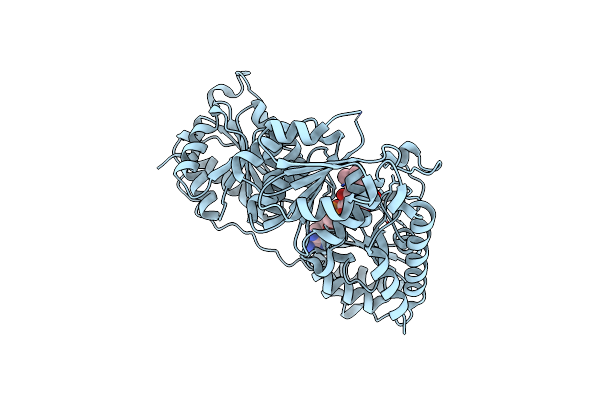
Crystal Structure Of Full-Length, Homohexameric 2-Oxoglutarate Dehydrogenase Kgd From Mycobacterium Smegmatis In Complex With Gara
Organism: Mycolicibacterium smegmatis mc2 155
Method: X-RAY DIFFRACTION Resolution:4.56 Å Release Date: 2023-08-16 Classification: OXIDOREDUCTASE Ligands: MG, CA, TPP |
 |
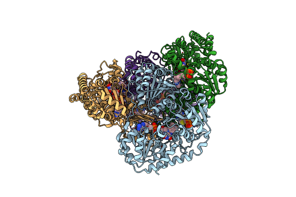
Crystal Structure Of The Homohexameric 2-Oxoglutarate Dehydrogenase Odha From Corynebacterium Glutamicum
Organism: Corynebacterium glutamicum atcc 13032
Method: X-RAY DIFFRACTION Resolution:2.46 Å Release Date: 2023-08-16 Classification: OXIDOREDUCTASE Ligands: EPE, COA, ACO, TPP, MG |
 |
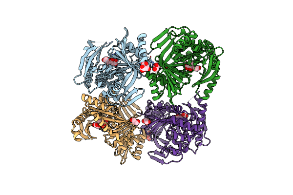
Single Particle Cryo-Em Structure Of The Homohexameric 2-Oxoglutarate Dehydrogenase Odha From Corynebacterium Glutamicum
Organism: Corynebacterium glutamicum atcc 13032
Method: ELECTRON MICROSCOPY Resolution:2.17 Å Release Date: 2023-08-16 Classification: OXIDOREDUCTASE Ligands: MG, ACO, TPP |
 |
Single Particle Cryo-Em Structure Of Homohexameric 2-Oxoglutarate Dehydrogenase Odha From Corynebacterium Glutamicum With Coenzyme A Bound To The E2O Domain
Organism: Corynebacterium glutamicum atcc 13032
Method: ELECTRON MICROSCOPY Resolution:2.17 Å Release Date: 2023-08-16 Classification: OXIDOREDUCTASE Ligands: MG, COA, ACO, TPP |
 |
Single Particle Cryo-Em Structure Of Homohexameric 2-Oxoglutarate Dehydrogenase Odha From Corynebacterium Glutamicum In Complex With The Product Succinyl-Coa
Organism: Corynebacterium glutamicum atcc 13032
Method: ELECTRON MICROSCOPY Release Date: 2023-08-16 Classification: OXIDOREDUCTASE Ligands: MG, ACO, TPP, SCA |
 |
Single Particle Cryo-Em Structure Of Homohexameric 2-Oxoglutarate Dehydrogenase Odha From Corynebacterium Glutamicum Following Reaction With The 2-Oxoglutarate Analogue Succinyl Phosphonate
Organism: Corynebacterium glutamicum atcc 13032
Method: ELECTRON MICROSCOPY Resolution:2.26 Å Release Date: 2023-08-16 Classification: OXIDOREDUCTASE Ligands: MG, ACO, QSP |
 |
Single Particle Cryo-Em Structure Of The Complex Between Corynebacterium Glutamicum Homohexameric 2-Oxoglutarate Dehydrogenase Odha And The Fha-Protein Inhibitor Odhi
Organism: Corynebacterium glutamicum atcc 13032
Method: ELECTRON MICROSCOPY Resolution:2.29 Å Release Date: 2023-08-16 Classification: OXIDOREDUCTASE Ligands: TPP, MG |
 |
Bovine Glutamate Dehydrogenase In Ternary Complex With The Allosteric Activators Adp And Leucine
Organism: Bos taurus
Method: X-RAY DIFFRACTION Resolution:2.45 Å Release Date: 2022-10-05 Classification: CYTOSOLIC PROTEIN Ligands: ADP, K, LEU |
 |
Organism: Bos taurus
Method: X-RAY DIFFRACTION Resolution:2.40 Å Release Date: 2022-10-05 Classification: CYTOSOLIC PROTEIN Ligands: ADP |
 |
Crystal Structure Of The S/T Protein Kinase Pkng From Corynebacterium Glutamicum In Complex With Amp-Pnp
Organism: Corynebacterium glutamicum (strain atcc 13032 / dsm 20300 / bcrc 11384 / jcm 1318 / lmg 3730 / ncimb 10025)
Method: X-RAY DIFFRACTION Resolution:2.20 Å Release Date: 2021-10-13 Classification: TRANSFERASE Ligands: MAP, MG |
 |
Crystal Structure Of The S/T Protein Kinase Pkng From Corynebacterium Glutamicum (Residues 130-433) In Complex With Amp-Pnp, Isoform 1
Organism: Corynebacterium glutamicum (strain atcc 13032 / dsm 20300 / bcrc 11384 / jcm 1318 / lmg 3730 / ncimb 10025)
Method: X-RAY DIFFRACTION Resolution:1.92 Å Release Date: 2021-10-13 Classification: TRANSFERASE Ligands: MG, MAP |