
Search Count: 26
 |
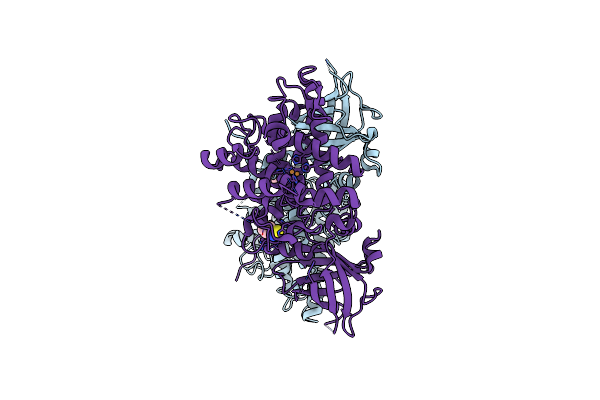
Organism: Plasmodium vivax, Synthetic construct
Method: X-RAY DIFFRACTION Resolution:1.60 Å Release Date: 2025-02-05 Classification: CELL INVASION Ligands: CA, NAG, SO4 |
 |
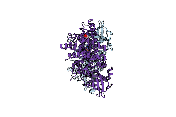
Organism: Plasmodium vivax, Synthetic construct
Method: X-RAY DIFFRACTION Resolution:1.50 Å Release Date: 2024-03-20 Classification: HYDROLASE Ligands: CA, NAG, SO4 |
 |
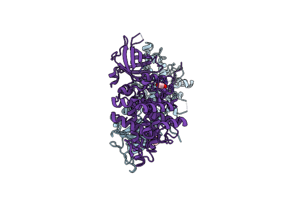
Organism: Plasmodium vivax, Synthetic construct
Method: X-RAY DIFFRACTION Resolution:1.54 Å Release Date: 2024-03-20 Classification: HYDROLASE Ligands: NAG, CA, SO4 |
 |
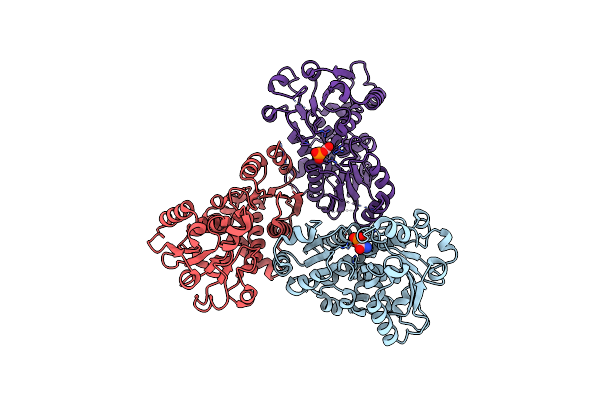
Organism: Plasmodium vivax, Synthetic construct
Method: X-RAY DIFFRACTION Resolution:1.77 Å Release Date: 2024-03-20 Classification: HYDROLASE Ligands: CA, NAG, SO4 |
 |
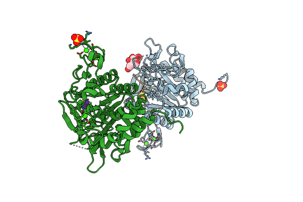
Structure Of The Catalytic Domain Of P. Vivax Sub1 (Triclinic Crystal Form) In Complex With Inhibitor
Organism: Plasmodium vivax, Synthetic construct
Method: X-RAY DIFFRACTION Resolution:1.51 Å Release Date: 2023-07-19 Classification: HYDROLASE Ligands: NAG, CA, SO4 |
 |
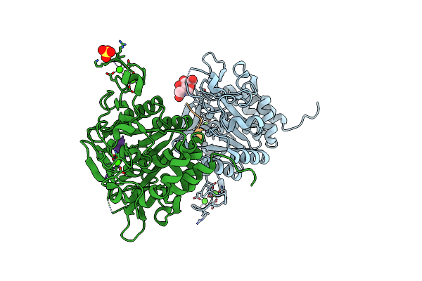
Structure Of The Catalytic Domain Of P. Vivax Sub1 (Triclinic Crystal Form)
Organism: Plasmodium vivax
Method: X-RAY DIFFRACTION Resolution:1.44 Å Release Date: 2023-07-19 Classification: HYDROLASE Ligands: CA, NAG, SO4 |
 |
Organism: Plasmodium vivax
Method: X-RAY DIFFRACTION Resolution:3.25 Å Release Date: 2023-07-19 Classification: HYDROLASE Ligands: CA |
 |
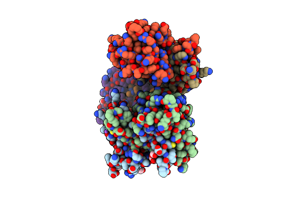
Organism: Severe acute respiratory syndrome coronavirus 2
Method: X-RAY DIFFRACTION Resolution:1.85 Å Release Date: 2022-06-22 Classification: RNA BINDING PROTEIN, Viral protein Ligands: GOL, ACT |
 |
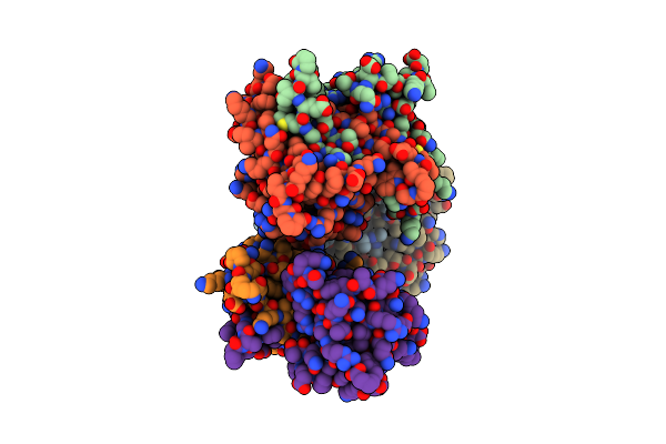
Crystal Structure Of Sars-Cov-2 Nucleocapsid Protein C-Terminal Domain Complexed With Chicoric Acid
Organism: Severe acute respiratory syndrome coronavirus 2
Method: X-RAY DIFFRACTION Resolution:1.73 Å Release Date: 2022-06-22 Classification: RNA BINDING PROTEIN/Viral Protein Ligands: GKP, PEG, GOL, NA, CL |
 |
Organism: Synthetic construct
Method: X-RAY DIFFRACTION Resolution:1.46 Å Release Date: 2021-05-19 Classification: UNKNOWN FUNCTION Ligands: EDO, PO4 |
 |
Organism: Unidentified
Method: X-RAY DIFFRACTION Resolution:1.55 Å Release Date: 2021-05-19 Classification: TRANSPORT PROTEIN Ligands: T44, EDO |
 |
Crystal Structure Of Tyrosinase Pvdp From Pseudomonas Aeruginosa Bound To Copper And Phenylthiourea
Organism: Pseudomonas aeruginosa (strain atcc 15692 / dsm 22644 / cip 104116 / jcm 14847 / lmg 12228 / 1c / prs 101 / pao1)
Method: X-RAY DIFFRACTION Resolution:2.40 Å Release Date: 2020-04-15 Classification: OXIDOREDUCTASE Ligands: URS, CU |
 |
Crystal Structure Of Tyrosinase Pvdp From Pseudomonas Aeruginosa Bound To Copper
Organism: Pseudomonas aeruginosa pao1
Method: X-RAY DIFFRACTION Resolution:2.70 Å Release Date: 2020-04-15 Classification: OXIDOREDUCTASE Ligands: CU, GOL |
 |
Organism: Pseudomonas aeruginosa (strain atcc 15692 / dsm 22644 / cip 104116 / jcm 14847 / lmg 12228 / 1c / prs 101 / pao1)
Method: X-RAY DIFFRACTION Resolution:2.11 Å Release Date: 2020-04-15 Classification: OXIDOREDUCTASE Ligands: GOL |
 |
Crystal Structure Of Truncated Aspartate Transcarbamoylase From Plasmodium Falciparum With Mutated Active Site (R109A/K138A) And N-Carbamoyl-L-Phosphate Bound
Organism: Plasmodium falciparum
Method: X-RAY DIFFRACTION Resolution:2.50 Å Release Date: 2018-09-26 Classification: TRANSFERASE Ligands: CP |
 |
Crystal Structure Of Truncated Aspartate Transcarbamoylase From Plasmodium Falciparum With Bound Inhibitor 2,3-Naphthalenediol
Organism: Plasmodium falciparum
Method: X-RAY DIFFRACTION Resolution:2.00 Å Release Date: 2018-02-21 Classification: TRANSFERASE Ligands: SO4, D48, DMS, MLI, GOL, NA, PEG |
 |
Crystal Structure Of The Complex Between Ppargamma Lbd And The Ligand Am-879
Organism: Homo sapiens
Method: X-RAY DIFFRACTION Resolution:2.69 Å Release Date: 2018-02-14 Classification: NUCLEAR PROTEIN Ligands: BKY |
 |
Crystal Structure Of Malate Dehydrogenase From Plasmodium Falciparum (Pfmdh)
Organism: Plasmodium falciparum (isolate 3d7)
Method: X-RAY DIFFRACTION Resolution:2.40 Å Release Date: 2018-01-31 Classification: OXIDOREDUCTASE Ligands: CIT |
 |
Crystal Structure Of Isoform 2 Of Purine Nucleoside Phosphorylase Complexed With Adenine
Organism: Schistosoma mansoni
Method: X-RAY DIFFRACTION Resolution:1.90 Å Release Date: 2017-10-11 Classification: TRANSFERASE Ligands: ADE, DMS |
 |
Crystal Structure Of Isoform 2 Of Purine Nucleoside Phosphorylase Complexed With Cytidine
Organism: Schistosoma mansoni
Method: X-RAY DIFFRACTION Resolution:2.10 Å Release Date: 2017-10-11 Classification: TRANSFERASE Ligands: CTN, SO4 |