
Search Count: 8
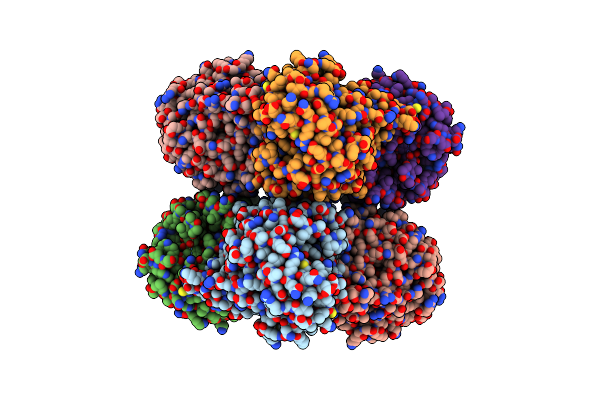 |
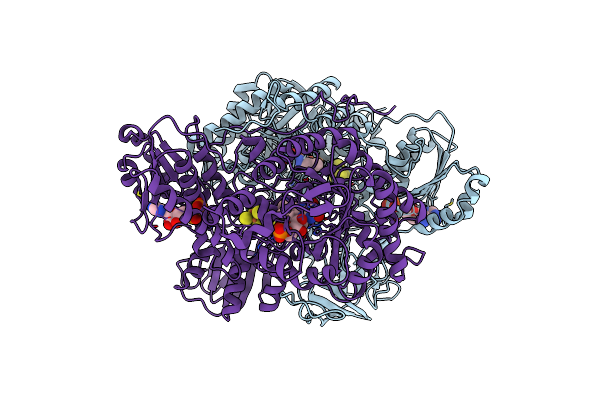
Crystal Structure Analysis Of Recombinant Rat Kidney Long-Chain Hydroxy Acid Oxidase
Organism: Rattus norvegicus
Method: X-RAY DIFFRACTION Resolution:2.30 Å Release Date: 2005-02-01 Classification: OXIDOREDUCTASE Ligands: FMN, ACY |
 |
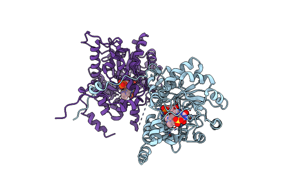
Crystallographic Study Of The Recombinant Flavin-Binding Domain Of Baker'S Yeast Flavocytochrome B2: Comparison With The Intact Wild-Type Enzyme
Organism: Saccharomyces cerevisiae
Method: X-RAY DIFFRACTION Resolution:2.30 Å Release Date: 2002-04-03 Classification: OXIDOREDUCTASE Ligands: HEM, FMN, MPD, PO4, PYR |
 |
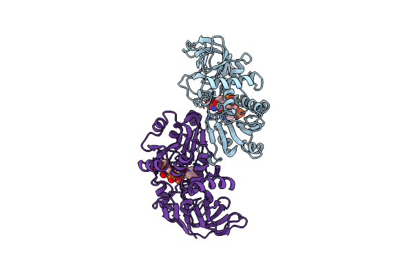
Crystallographic Study Of The Recombinant Flavin-Binding Domain Of Baker'S Yeast Flavocytochrome B2: Comparison With The Intact Wild-Type Enzyme
Organism: Saccharomyces cerevisiae
Method: X-RAY DIFFRACTION Resolution:2.50 Å Release Date: 2002-04-03 Classification: OXIDOREDUCTASE Ligands: FMN, EDO |
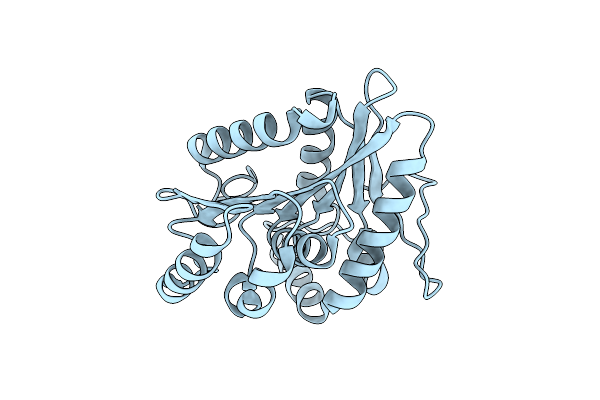 |
Crystal Structure Of The C123S Mutant Of Dienelactone Hydrolase (Dlh) Bound With The Pms Moiety Of The Protease Inhibitor, Phenylmethylsulfonyl Fluoride (Pmsf)
Organism: Pseudomonas putida
Method: X-RAY DIFFRACTION Resolution:2.50 Å Release Date: 2000-12-13 Classification: HYDROLASE |
 |
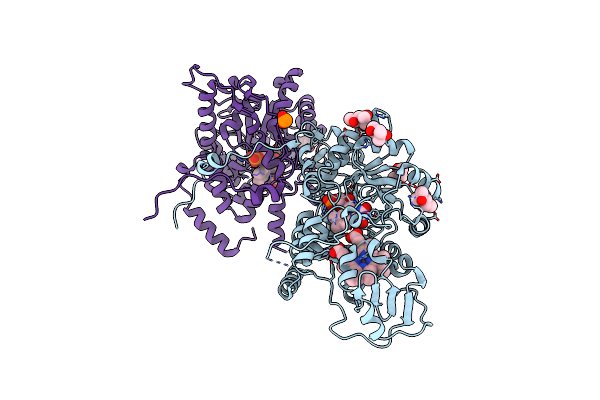
Structural And Biochemical Characterization Of Recombinant Wild Type Trimethylamine Dehydrogenase From Methylophilus Methylotrophus (Sp. W3A1)
Organism: Methylophilus methylotrophus
Method: X-RAY DIFFRACTION Resolution:2.20 Å Release Date: 1999-12-22 Classification: OXIDOREDUCTASE Ligands: SF4, FMN, ADP |
 |
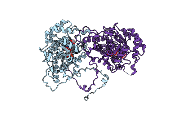
Structural And Biochemical Characterization Of Recombinant C30A Mutant Of Trimethylamine Dehydrogenase From Methylophilus Methylotrophus (Sp. W3A1)
Organism: Methylophilus methylotrophus
Method: X-RAY DIFFRACTION Resolution:2.20 Å Release Date: 1999-12-22 Classification: OXIDOREDUCTASE Ligands: SF4, FMN, ADP |
 |
Organism: Saccharomyces cerevisiae
Method: X-RAY DIFFRACTION Resolution:2.75 Å Release Date: 1999-05-24 Classification: OXIDOREDUCTASE Ligands: FNS |
 |
Crystal Structure Of Escherichia Coli Quinone Oxidoreductase Complexed With Nadph
Organism: Escherichia coli
Method: X-RAY DIFFRACTION Resolution:2.20 Å Release Date: 1995-06-03 Classification: OXIDOREDUCTASE Ligands: SO4, NDP |